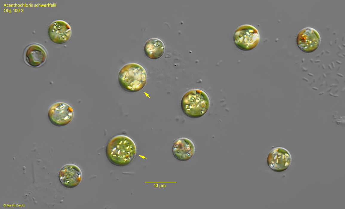
I find Acanthochloris scherffelii only very rarely and always only when specimens have settled on the floating coverslip. In fresh samples, the small cells are difficult to identify. So far, I have only been able to identify Acanthochloris scherffelii in samples from the Simmelried in this way.
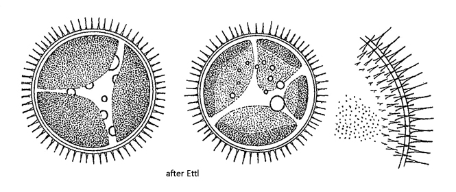
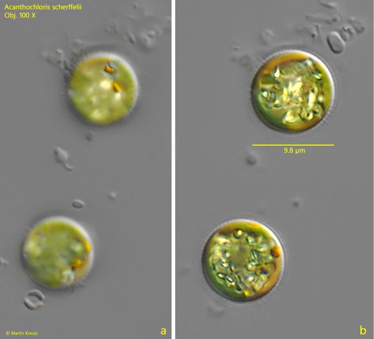
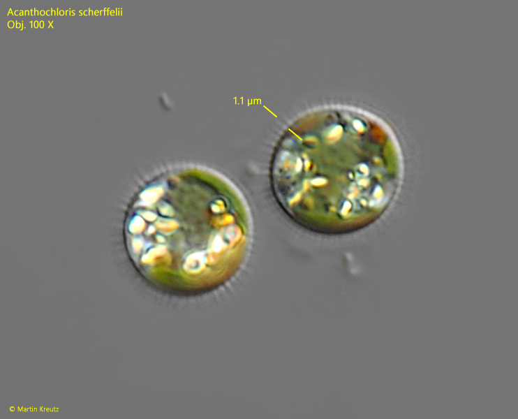
The main characteristic of Acanthochloris scherffelii is a fringe of needle-shaped spines that cover the cell wall (s. figs. 1 and 3). However, these can only be seen at high magnification. In specimens with well-developed spines, these were 1.0–1.1 µm long in my population (s. fig. 3). However, I was able to determine that the spines can also be significantly shorter and then difficult to recognize. Many cells in my population contained conspicuous orange oil droplets. Although Ettl (1978) describes that the cells often contain oil droplets, he does not mention the conspicuous coloration. The cells in my population had a diameter of 7.6–9.8 µm, which fits well with Ettl’s data (8–14 µm).
Fig. 1:Acanthochloris scherffelii. D = 7.6–9.4 µm. Several specimens which settles on a floating coverslip. Note the delicate, needle-shaped spines covering the cell wall (arrows). Obj. 100 X.
Fig. 2 a-b:Acanthochloris scherffelii. D = 9.8 µm. Two focal planes of of two larger cells. The cells contain some orange oil droplets. Obj. 100 X.
Fig. 3:Acanthochloris scherffelii. D = 7.7–7.8 µm. The delicate spines of these two cells have a length of 1.0–1.1 µm. Obj. 100 X.