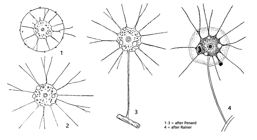
cell almost spherical, variable in shape, covered with gelatinous layer
cell diamter 16–30 µm
cell on a hollow stalk, sometimes stalk absent
base of stalk root-like
filopodia with granules, sometimes branched
serveral contractile vacuoles
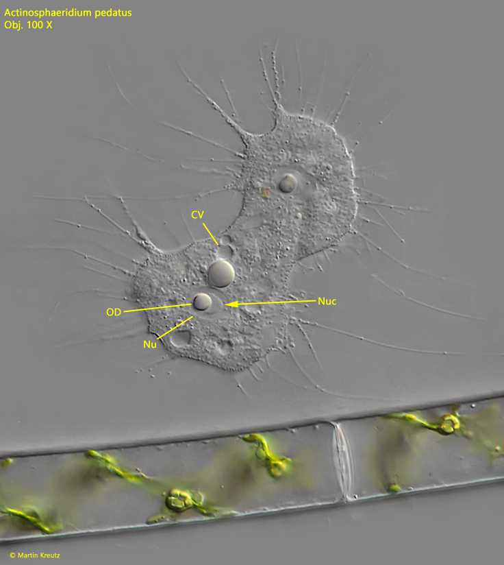
nucleus with central nucleolus and oil droplets
Actinosphaeridium pedatus
I have only found Actinosphaeridium pedatus in the Simmelried so far. The species is rarely found in fresh samples because it sits on algae and detritus particles and is difficult to detect in between. However, the species likes to settle on floating cover glasses and can then be observed in isolation.
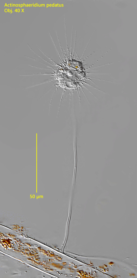
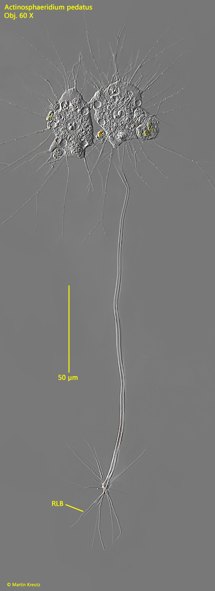
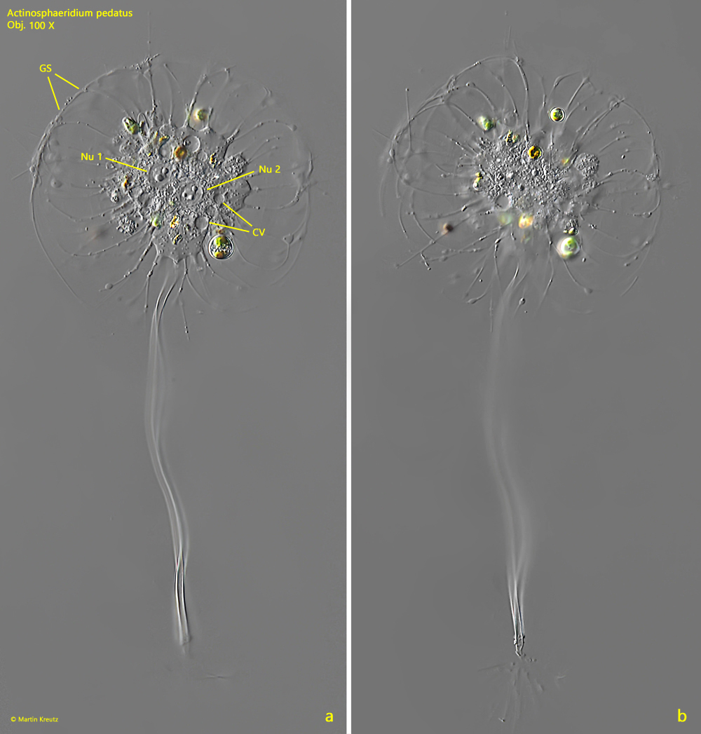
In my population, most of the specimens had formed a stalk. This can vary in length. Mostly between 100–150 µm. The diameter of the hollow stalk is 1.6–2 µm. At the base, the stalk forms root-shaped fibers with which it attaches to the substrate (s. fig. 2). The cells itself had a diameter of 20–26 µm in my population. The cells are surrounded by an invisible gelatinous sheath. Its shape and dimensions can only be recognized by adhering bacteria or when the filopodia are bent after the coverslip has been placed. They only bend outside the gelatinous sheath. Inside they are stabilized by it (s. fig. 3 a-b). I was able to observe oil drops around and in the nucleus in all the specimens I observed (s. fig. 4). These are very conspicuous, but were not mentioned by earlier authors. They seem to be species-specific. The fact that the oil droplets are also located inside the nucleus can be seen in heavily squashed cells. The oil droplet remains in the nucleus and does not move away with the spreading cytoplasm. There is also a nucleolus in the center of the nucleus, which is also not mentioned by earlier authors (s. fig. 4). It seemed to me that the oil droplet and the nucleolus were associated.
Fig. 1:Actinosphaeridium pedatus. L = 166 µm (with stalk). Total view of a specimen attached to a dead filamentous alga. Obj. 40 X.
Fig. 2:Actinosphaeridium pedatus. L = 245 µm (with stalk). A slightly squashed specimen during cell division. The specimen was attached to a floating coverslip with the root-like base of the stalk (RBL). Note the refractive oil droplets in the nuclei. Obj. 60 X.
Fig. 3 a-b: Actinosphaeridium pedatus. L = 102 µm (with stalk). Two focal planes of a slightly squashed specimen with two nuclei (Nu 1, Nu 2) after placing the coverslip. Due to the mechanical stress, the filopodia outside of the gelatine sphere have been bent, revealing the extent of this sphere. In this specimen, the diameter of the cell including the gelatinous sphere is 52 µm. Obj. 100 X.
Fig. 4: Actinosphaeridium pedatus. A squashed specimen without stalk in cell division. Note the nucleolus (Nuc) and the oil droplet (DP) in the nucleus (Nu). The oil droplet is located in the nucleus and not outside, because it does not move away from the nucleus under coverslip pressure. Obj. 100 X.