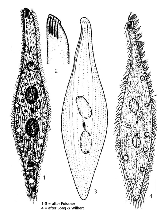
body slender lanceolate, sigmoid, slightly contractile
length 150–450 µm, width 40–60 µm
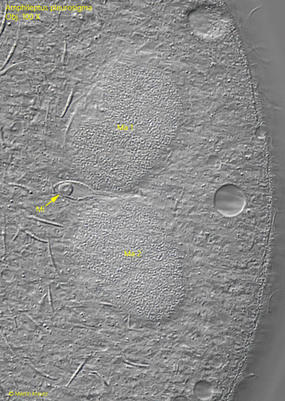
two ellipsoidal macronuclei with one micronucleus in between
micronucleus in a membraneous sac (funiculus)
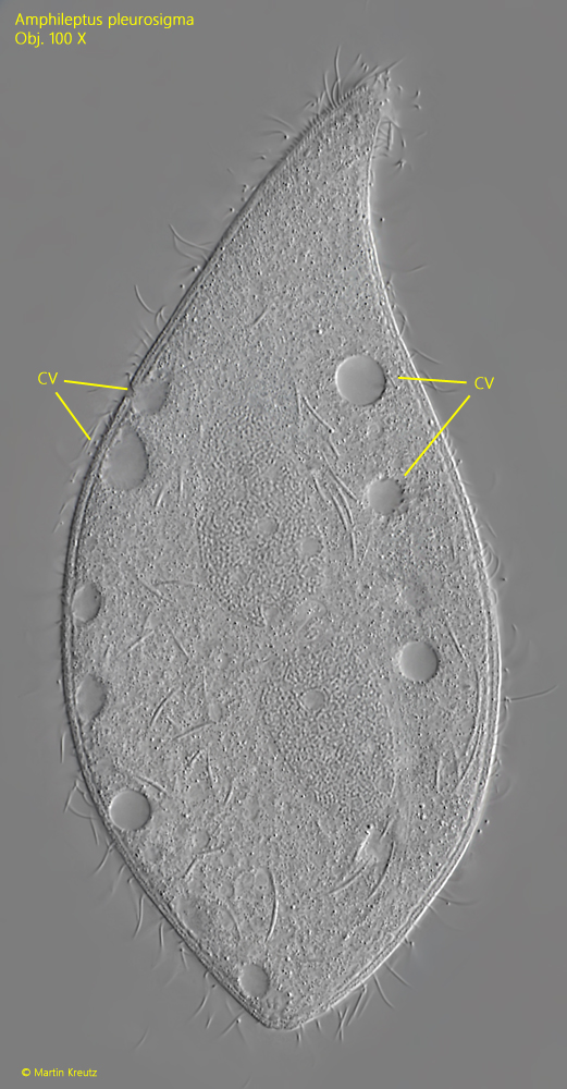
two marginal rows of contractile vacuoles
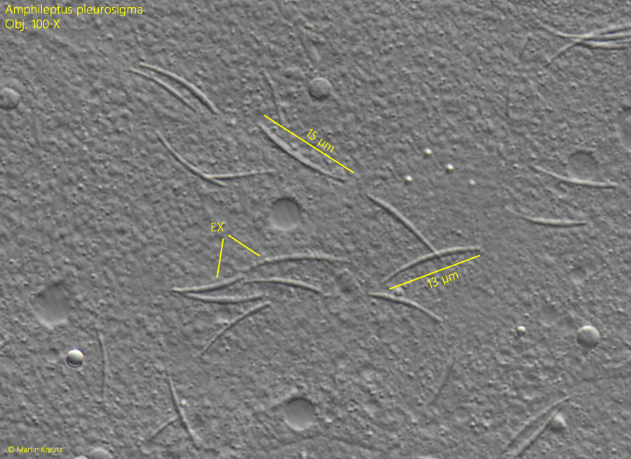
bundle of extrusomes in apical end
extrusomes curved, tusk-shaped, 10-15 µm long
several extrusomes scattered in cytoplasm
right side with 25–35 longitudinal rows of cilia
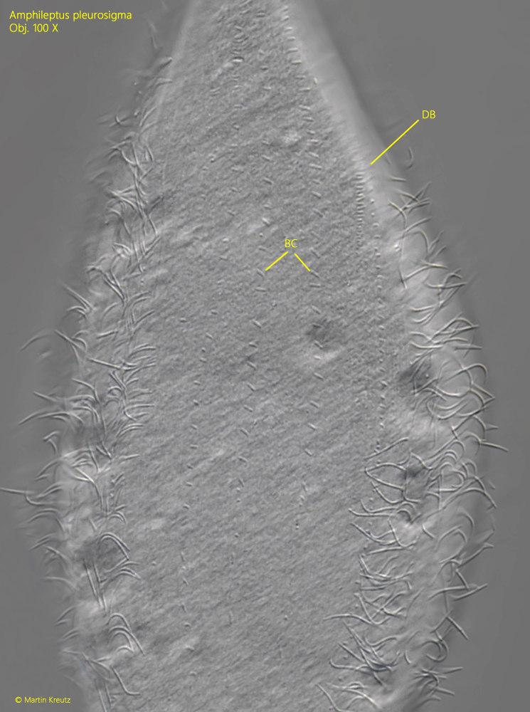
left side with 4–6 rows of bristl-like, short cilia
oral cleft only recognizable during feeding
Amphileptus pleurosigma
I find Amphileptus pleurosigma very frequently, especially in the Purren pond, a body of water with lots of fallen leaves. Amphileptus pleurosigma can be found here practically all year round.
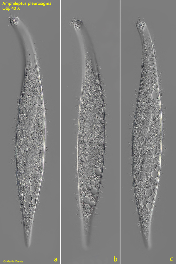
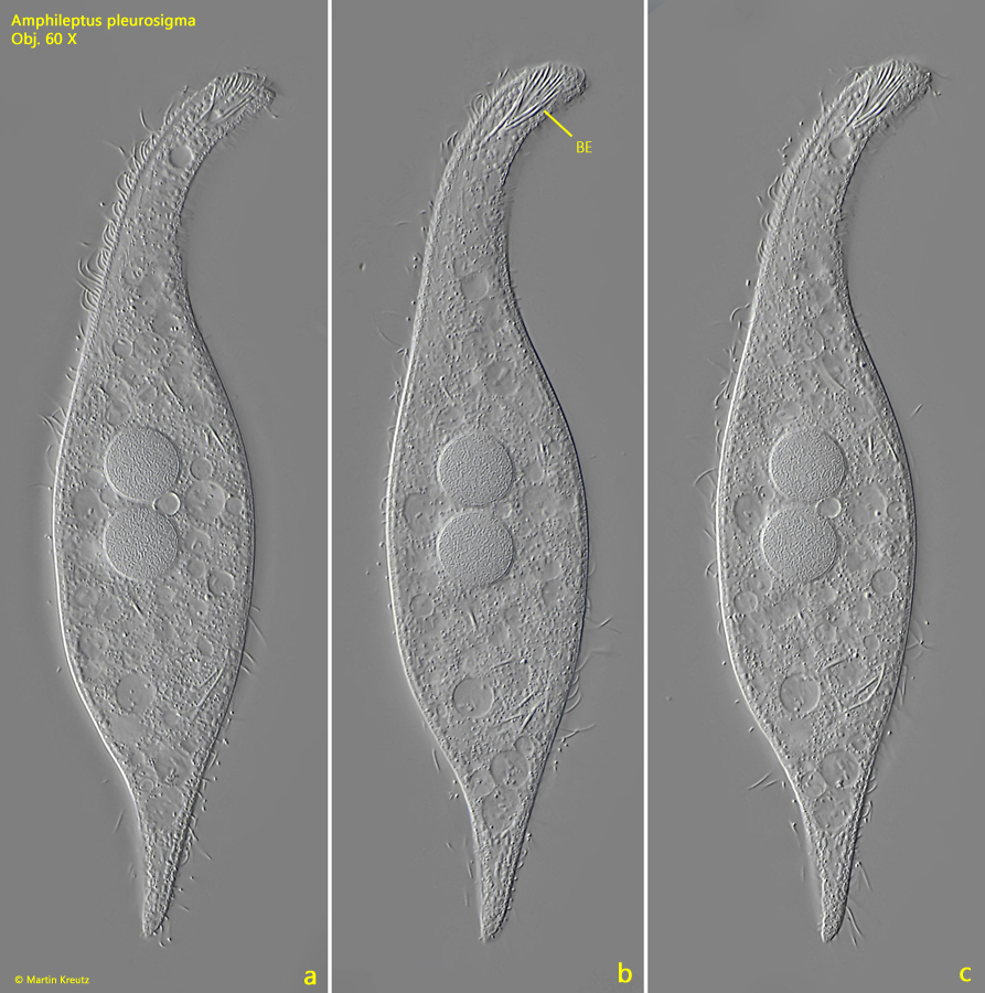
The specimens in my population were between 200–350 µm long and swim quite fast. In the free-swimming state, the body is very slender and slightly sigmoid. The bundle of curved extrusomes at the apical end is clearly visible. Only in slightly squashed specimens can one recognize that further extrusomes are distributed in the cytoplasm (s. fig. 2 a-c).
Amphileptus pleurosigma is a pleurostomatid ciliate. Some genera of this order (e.g. Litonotus or Loxophyllum) have a bilateral structure with a ciliated right side and a rudimentarily ciliated left side. Often the left side is also naked. In the case of Amphileptus pleurosigma, there are only 4–6 rows of very short, bristle-like cilia on the left side (s. fig. 5).
The mouth opening of Amphileptus pleurosigma is not located where the apical bundle of extrusomes is, but is a ventral slit in the anterior third, which can only be seen when feeding. Amphileptus pleurosigma prefers to feed on peritrichous ciliates.
The nuclear apparatus of Amphileptus pleurosigma consists of two macronuclei and a micronucleus in between. This is enclosed in a clearly visible membranous sac called the funiculus (s. fig. 6). Together with the curved extrusomes, which have the shape of a tusk, this funiculus is an important identifying feature.
Fig. 1 a-c:Amphileptus pleurosigma. L = 342 µm. A freely swimming specimen. Obj. 40 X.
Fig. 2 a-c:Amphileptus pleurosigma. L = 185 µm. A slightly squashed specimen. In the apical end a bundle of curved extrusomes (BE) is visible. Obj. 60 X.
Fig. 3:Amphileptus pleurosigma. A squashed specimen for visualisation of the two marginal rows of contractile vacuoles (CV). Obj. 100 X.
Fig. 4:Amphileptus pleurosigma. The border between the right and left side (arrows). The right side is ciliated, while the left side is almost naked. Obj. 100 X.
Fig. 5:Amphileptus pleurosigma. A squashed specimen from left. On this side only a few rows of short, bristle-like cilia (BC) are present. DB = part of the dorsal brush. Obj. 100 X.
Fig. 6:Amphileptus pleurosigma. The two macronuclei (Ma 1, Ma 2) and the micronucleus (Mi) in a membraneous sac (= funiculus). Obj. 100 X.
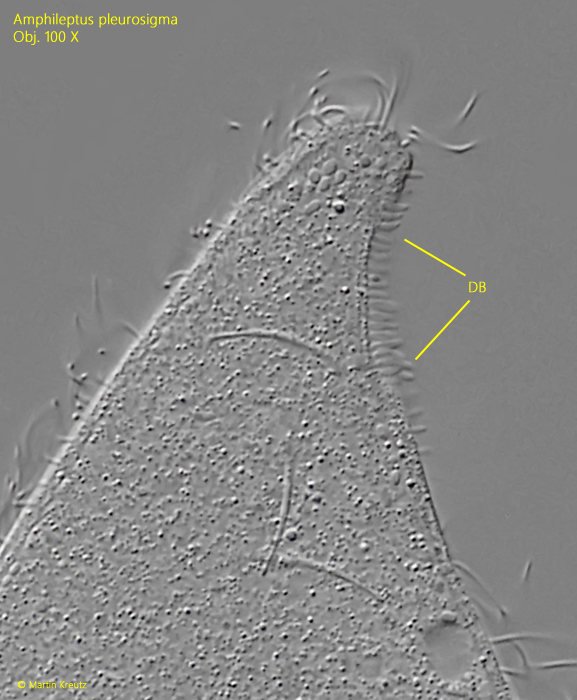
Fig. 7:Amphileptus pleurosigma. Focal plane on the dorsal brush (DB). Obj. 100 X.
Fig. 8:Amphileptus pleurosigma. The curved extrusomes are 13–15 µm long and tusk-shaped. Obj. 100 X.
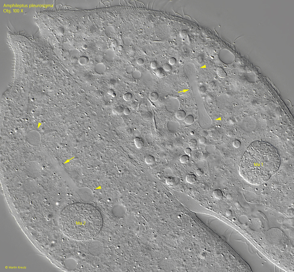
Fig. 9:Amphileptus pleurosigma. A squashed pair in conjugation during the process of meiotic maturation division of the micronuclei. Between the already separated chromosomes (arrowheads) the elongated spindle apparatus (arrow) is visible. The macronuclei (Ma 1, Ma 2) are already condensed. After conjugation, they are broken down and newly formed from the exchanged micronuclei. Obj. 100 X.