Amphitrema wrightianum has so far only been found by me in July 2017 in the Sima Moor and in August 2025 in the Lauchsee Moor. In the locations in my vicinity, I have not been able to detect this testate amoeba so far.
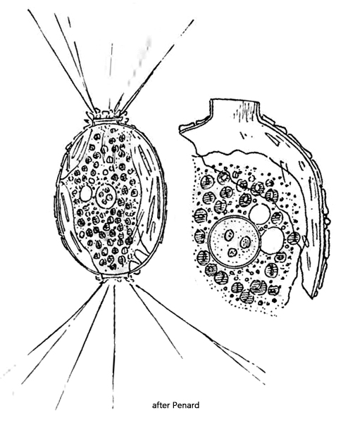
Amphitrema wrightianum is easy to identify by the oval shell with two opposite apertures, which have a very short neck. The cytoplasm of the amoeba is green-colored due to numerous symbiotic algae (s. fig. 2). They lie so densely that both the nucleus and the contractile vacuole are difficult to recognize. The symbiotic algae themselves do not seem to have been examined in detail by previous authors. They can only be clearly seen when the shell is strongly compressed and the algae are then released (s. fig. 3). According to my observations, they are about 10–11 µm long, ellipsoid, oval, or kidney-shaped. Their shape is often asymmetrical. Each possesses two pyrenoids and (probably) two chloroplasts. In the center of each algal cell, there are starch grains that fluoresce strongly in DIC. I could not detect a nucleus in the symbiotic algae.
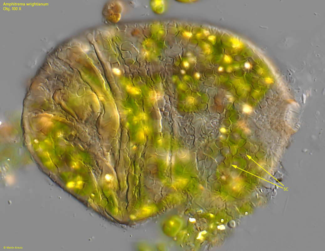
The shell consists of an organic material in which xenosomes are embedded or glued. As a result, the shell is often opaque. Xenosomes include mineral grains, diatom shells, or flat scales. In the organic matrix of the shell of my population, flat, oval scales were embedded, measuring 3-4 µm in length (s. fig. 4). Potentially, these are the scales of shells from other amoebae.
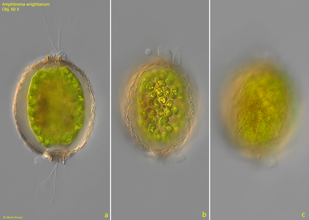
Fig. 1 a-c:Amphitrema wrightianum. L = 93 µm (of shell). Three focal planes of a specimen found in August 2025 in the Lauchsee Moor. Obj. 60 X.
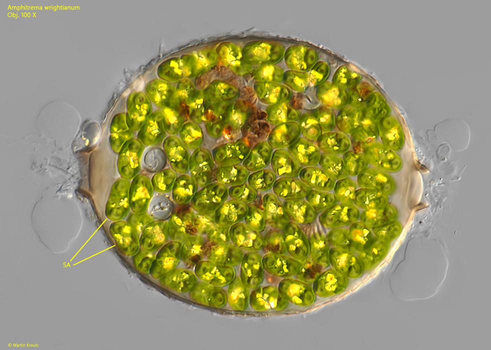
Fig. 2:Amphitrema wrightianum. In the squashed specimen the symbiotic algae (SA) scattered in the cytoplasm become visible. Obj. 100 X.
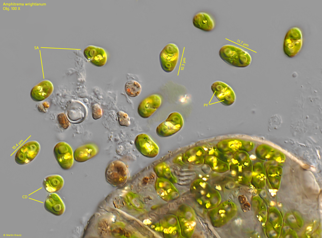
Fig. 3:Amphitrema wrightianum. The relaesed symbiotic algae (SA) in a strongly squashed specimen. They have a length of about 10–11 µm and two pyrenoids (PY) per cell. The bright, yellowish granules in the center of the cells are starch grains. One cell is in the process of cell division (CD). Each daughter cell has only one pyrenoid. Obj. 100 X.
Fig. 4:Amphitrema wrightianum. Focal plane on the surface of the shell. The xenosomes of this specimen are scales (SC) with a more or less oval shape and a length of 3–4 µm. Obj. 100 X.