Amphizonella violacea Greeff, 1866

Most likely ID: n.a.


Synonym: n.a.


Sampling location: Moss


Phylogenetic tree: Amphizonella violacea


Diagnosis: 

  • body globular, very slow movement
  • pseudopodia hyaline and clear
  • diameter 125–250 µm
  • soft and flexible shell, thickness 3–23 µm
  • pseudostome irregularly shaped
  • cytoplasm filled with numerous violet vesicles
  • one ellipsoid or globular nucleus, diameter up to 40 µm
  • about 20 contractile vacuoles near surface
Amphizonella-violacea
Amphizonella violacea

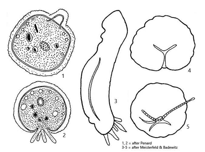
So far, I have found Amphizonella violacea exclusively in mosses on roofs. After moistening the moss samples with collected rainwater, the first excysted specimens were found after 2 days. In 2006, Meisterfeld and Badewitz published a redescription of Amphizonella violacea. The authors also found their specimens in mosses on roofs. However, other authors such as Siemensma (s. link below) and Glück (2007) report that Amphizonella violacea can also be found in Sphagnum bogs.


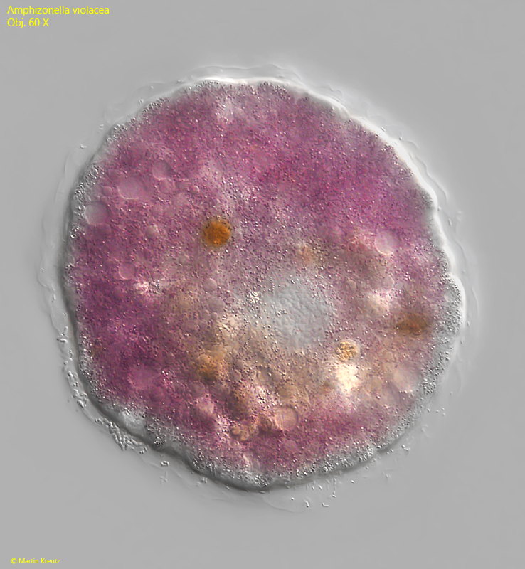
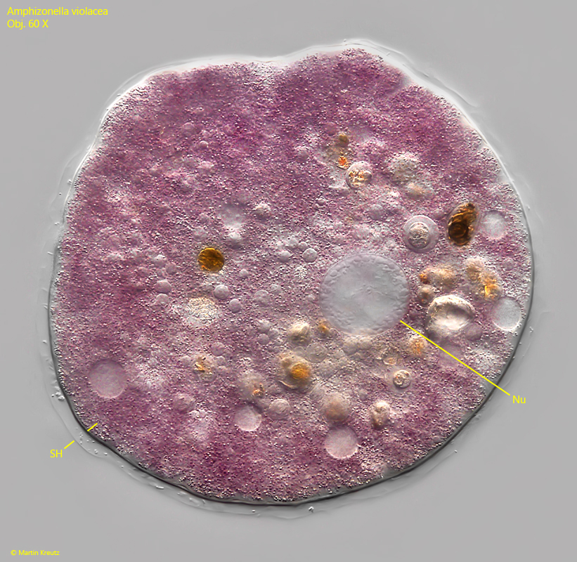
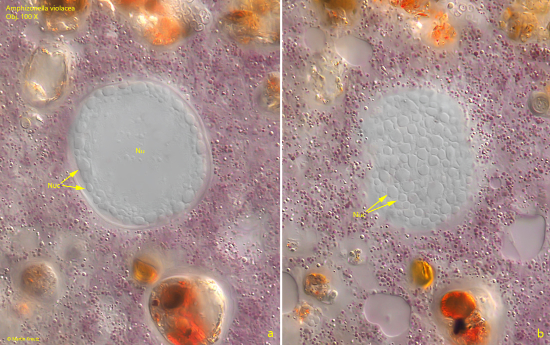
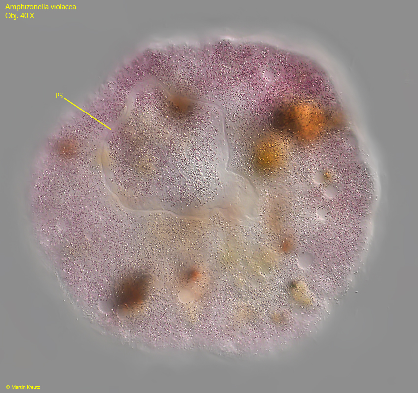
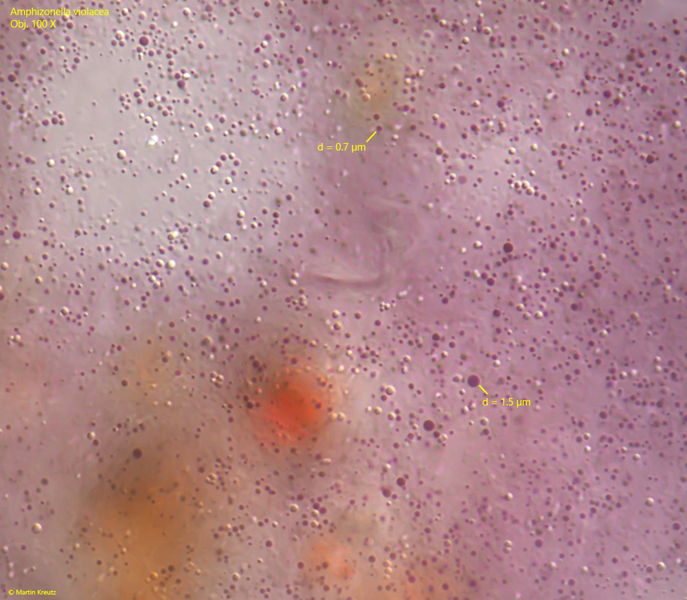
At low magnifications, the rounded specimens are easy to overlook because they often appear black and look like lifeless detritus particles. Only at higher magnification and with slight coverslip pressure can the distinct violet coloration of Amphizonella violacea be recognized. This coloration is produced by large amounts of violet-stained vesicles with a diameter of 0.1–2 µm, which are distributed throughout the cytoplasm (s. fig. 6).


The specimens of Amphizonella violacea moved very little and then only very slowly. Most of the time, the body was rounded or knobby. The rounded specimens had a diameter of 160–230 µm. The specimens phagocytized large amounts of detritus, algae, and also mineral grains. Everything was phagocytized completely non-specifically.


In all specimens of my population, I could only identify one nucleus. This was ellipsoid or round in shape with a diameter of 35–40 µm. Numerous, flake-shaped nucleoli were visible in the periphery (s. fig. 3 a-b). This corresponds with the observations of Meisterfeld & Badewitz (2006), Glück (2007), and Siemensma (s. link below). Only Penard (1902) reported finding specimens with 2 or 3 nuclei. In slightly compressed specimens, numerous contractile vacuoles can be seen, all located just beneath the cell membrane.


The shell of Amphizonella violacea consists of a soft and flexible, chitinous layer. In some specimens, I was able to observe layering. The shell was either colorless or distinctly yellow-brown (s. figs 5 and 7). In some cases, I also observed how the shell clearly detached from the cell body, creating blister-like elevations (s. fig. 7). This detachment of the shell may be caused by the movement of the cell body, which the shell cannot adapt to. However, this phenomenon illustrates that the shell lies loosely against the plasma body. The thickness of the shell in Amphizonella violacea seems to be subject to some variability. I measured a range of 1–5 µm. Meisterfeld and Badewitz report a thickness of up to 25 µm. The shell can be covered by a gelatinous layer, in which bacteria are often found. However, I could not detect this layer in all specimens, which corresponds with the observations of Siemensma (s. link below).


Amphizonella violacea is difficult to distinguish from the very similar, also violet-colored species Zonomyxa violacea. Meisterfeld and Badewitz (2006) cite the following essential differences:


Amphiozella violacea has only one nucleus, while Zonomyxa violacea has 4–32
– a gelatinous layer over the shell is absent in Zonomyxa violacea
Zonomyxa violacea is said to have a thinner shell than Amphizonella violacea
– the habitat of Zonomyxa violacea is Sphagnum bogs, while Amphizonella violacea lives in xerophilous mosses (on roofs, walls, or trees) with strongly fluctuating water content


However, in my opinion, some of these characteristics are very unspecific. The habitats overlap, as Amphizonella violacea has also been found in Sphagnum bogs. The thickness of the shell is very variable in both species, and here too the measurements overlap. Finally, the gelatinous sheath can also be absent in Amphizonella violacea. The number of nuclei remains the most reliable characteristic. Amphizonella violacea always seems to be uninucleate.


More images and information on Amphizonella violacea: Ferry Siemensma-Microworld-Amphizonella violacea

Amphizonella-violacea

Fig. 1: Amphizonella violacea. D = 158 µm. A rounded, resting specimen. Obj. 60 X.

Amphizonella-violacea

Fig. 2: Amphizonella violacea. D = 158 µm. In a slightly squashed specimen the single, globular nucleus (Nu) is visible. SH = shell. Obj. 60 X.

Amphizonella-violacea

Fig. 3 a-b: Amphizonella violacea. Two focal planes of the nucleus (Nu) with numerous peripher nucleoli (Nuc). The nucleus has a diameter of 40 µm. Obj. 100 X.

Amphizonella-violacea

Fig. 4: Amphizonella violacea. D = 220 µm. Ventral view on the irregularly shaped pseudostome (PS). Obj. 40 X.

Amphizonella-violacea

Fig. 5: Amphizonella violacea. The chitinoid shell (SH) of this specimen has a thickness of about 3 µm. A gelatinous sheath is absent. CV = contractile vacuoles. Obj. 100 X.

Amphizonella-violacea

Fig. 6: Amphizonella violacea. The violet vesicles scattered in the cytoplasm have a diameter of 0.5 to 1.5 µm. Obj. 100 X.

Amphizonella-violacea

Fig. 7: Amphizonella violacea. In some specimens the shell was colored yellowish-brown and detached from the cell surface in a blister-shape (arrows). CV = contractile vacuoles. Obj. 60 X.