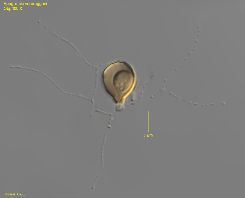
shell pyriform or retort shaped, sometimes irregular
shell more or less mirror-symmetrical
shell with distinct neck (sometimes absent)
shell often orange-brown due to iron precipitate
length 8.5–12.7 µm
anastoming network of granulated pseudopodia
Apogromia verbrugghei
I find Apogromia verbrugghei exclusively in the Simmelried. The specimens are hard to find in fresh samples due to its small size. However, Apogromia verbrugghei likes to settle on the floating coverslips and is then easy to observe.
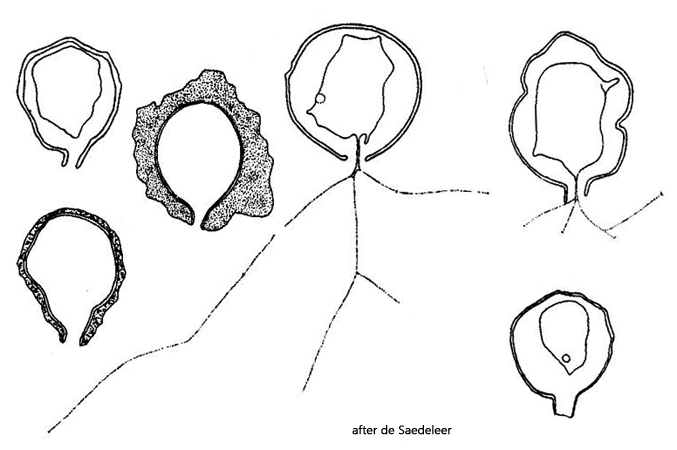
De Saedeleer (1934) originally found Apogromia verbrugghei in salt water in the harbor of Blankenberge on the Flemish coast and described it for the first time. As the specimens came from salt water, he was unable to detect a contractile vacuole. However, he mentions the high variability of the species and also illustrated the different forms (s. drawings above).
The specimens of my population were 8.0–12.3 µm long and the shells were always orange-brown. I found both symmetrically shaped specimens without a neck (s. figs 1) and asymmetrically shaped specimens with a neck (s. figs. 2 and 3). The nucleus is located at the posterior end and has a central nucleolus (s. fig. 2). The contractile vacuole was mostly found in the middle of the cell or in the anterior third (s. fig. 2). I have only rarely found specimens with a widely extended network of granulated pseudopodia (s. fig. 1). Mostly there were 3–4 very long but little branched pseudopodia (s. fig. 3). The cell almost never completely fills the shell. I could not observe any division.
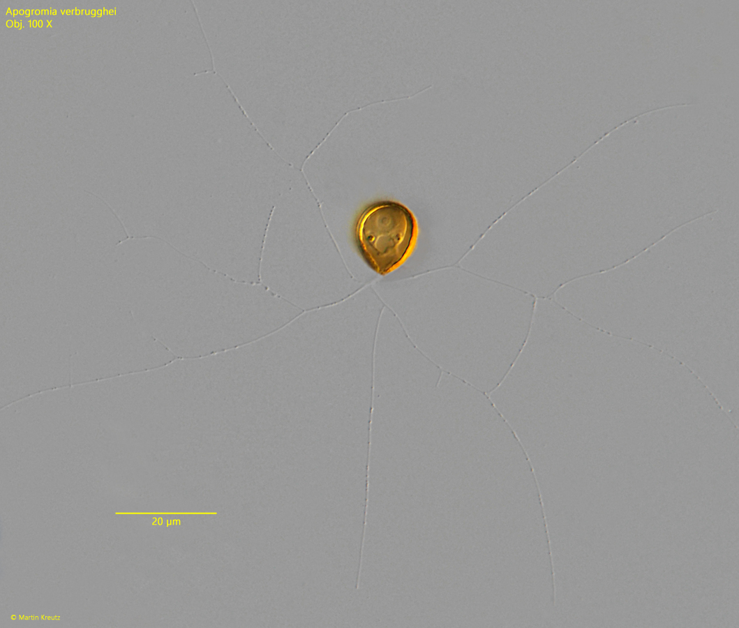
Fig. 1:Apogromia verbrugghei. L = 12.3 µm. A specimen with a symmetrical, pyriform shell but without a distinct neck. The granulated pseudopodia form an extended network. Obj. 100 X.
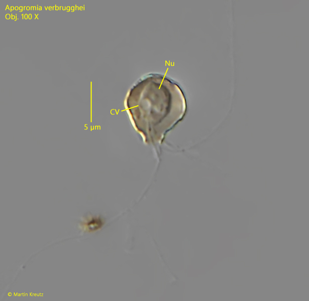
Fig. 2:Apogromia verbrugghei. L = 8.1 µm. A more irregularly shaped specimen with a neck. The nucleus (NU) is located posterior and the contractile vacuole (CV) in mid-body. Obj. 100 X.
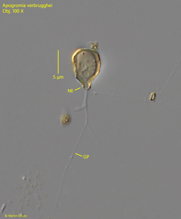
Fig. 3:Apogromia verbrugghei. L = 8.7 µm. A second irregularly shaped specimen with a distinct nec (NE). GP = granulated pseudopodia. Obj. 100 X.
Fig. 4:Apogromia verbrugghei. L = 9.6 µm. A symmetrically shaped specimen with an indistinct neck and an retracted network of pseudopodia. Obj. 100 X.