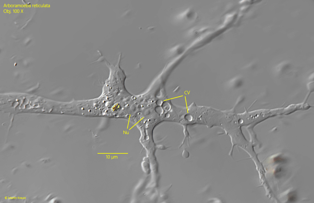
numerous contractile vacuoles distribute over network
cytoplasm with food particles and crystals
No drawings from previous autors available.
I regularly find Arboramoeba reticulata in detritus flakes from the Simmelried. The specimens burrow into the detritus flakes and are therefore difficult to discover. Only when the detritus flakes are slightly squashed under the coverslip the specimens can be recognized by the extensively branched pseudopodia.
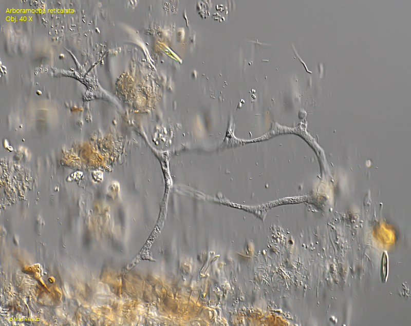
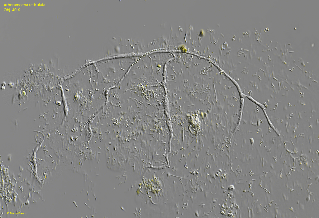
Arboramoeba reticulata was only scientifically described in 2015 by Geisen, Bass & Berney. Characteristic of this amoeba are the slender and elongated pseudopodia, which can anastomose and from which further, highly branched extensions branch off. At their ends, fine, straight filopodia arise.
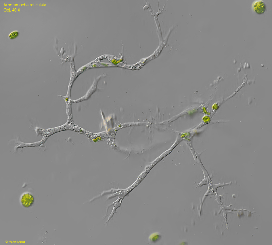
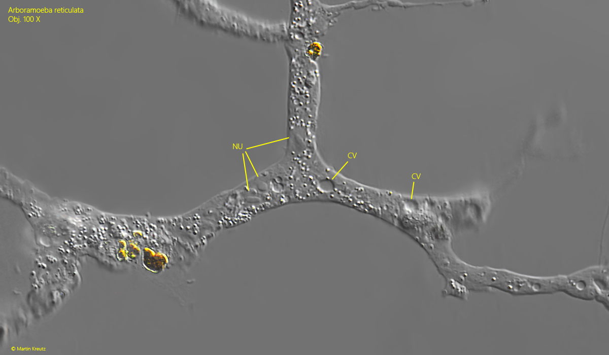
The pseudopodia form a three-dimensional network within the detritus flakes, which in some cases penetrates the entire detritus flake. Only very rarely have I found almost exposed specimens (s. fig. 3). Numerous nuclei and contractile vacuoles are scattered in the cytoplasm. The nuclei possess a central nucleolus that can be round, oval, or also loaf-shaped. In addition, highly refractive granules and phagocytosed food are present. The food consists of bacteria, flagellates, small algae, and cyanobacteria. In the pseudopodia I was able to observe a distinct plasma streaming that ran in both directions.
The reticulose networks of the specimens in my population had a diameter of about 200–300 µm. I find most specimens in winter and spring.
Fig. 1:Arboramoeba reticulata. D = 240 µm. A specimen in a detritus flake. Obj. 40 X.
Fig. 2:Arboramoeba reticulata. D = 290 µm. A second specimen in a gelatinous detritus flake. Obj. 40 X.
Fig. 3:Arboramoeba reticulata. D = 310 µm. A third specimen slightly covered with bacteria and detritus. Obj. 40 X.
Fig. 4:Arboramoeba reticulata. D = 240 µm. A part of the branched pseudopodia. The nuclei (Nu) have an asymmetrical shaped nucleolus in the center. Many contractile vacuoles are scattered over the pseudopodia. Obj. 40 X.
Fig. 5:Arboramoeba reticulata. The nuclei (Nu) and contractile vacuoles (CV) in a second specimen. Obj. 100 X.