Aspidiophorus oculifer Kisielewski, 1981

Most likely ID: n.a.


Synonym: n.a.


Sampling location: Mühlweiher Litzelstetten, Simmelried


Phylogenetic tree: Aspidiophorus oculifer


Diagnosis: 

  • body small sized, stocky, sole-shaped
  • length 115–135 µm
  • head 5-lobed with 2 lateral ocelli or ocellar granules
  • hypostomium weakly developed
  • pharynx slightly dumbbel-shaped
  • dorsal 17–22 longitudinal rows of leal-shaped scales
  • dorsal scales 2.5–4 µm long, distal end tapered
  • ventral field with 9–10 rows of small keeled scales
  • two spined terminal scales ventrally
  • adhesive tubes short, end in notches
Aspidiophorus-oculifer
Aspidiophorus oculifer
Aspidiophorus oculifer is described as a rare species (Schwank, 1990) and I have so far only found 2 specimens. The first was among floating aquatic plants in the Mühlweiher Litzelstetten and the second specimen in the mud of the Simmelried.
 
My specimens were slightly smaller at 100 µm and 111 µm than those reported by Schwank (115–135 µm). A key identification feature are the ocelli (also called pseudocells), which can sometimes also consist of an aggregation of ocellar granules, as was the case with my specimens (s. fig. 2). The dorsal scales of Aspidiophorus oculifer are very small and their shape is difficult to discern due to their overlapping arrangement. Only when the scales can be detached by shifting the cover glass does their shape, similar to a birch leaf, become visible (s. fig. 4). In my specimens, the dorsal scales were 3.6-3.9 µm long.
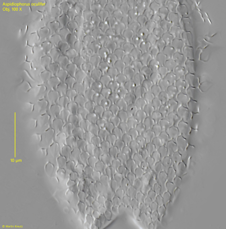
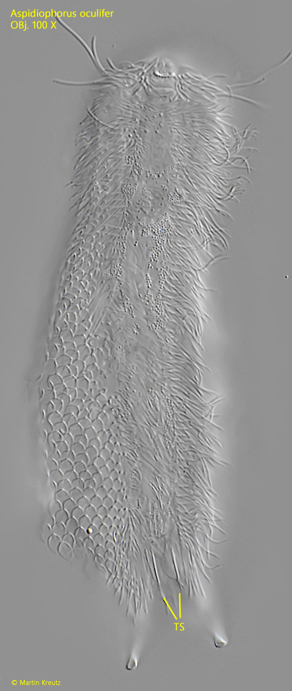
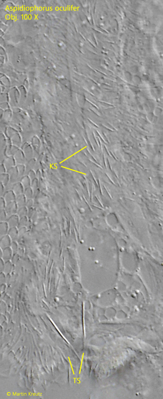
The narrow, keeled scales on the ventral side could only be recognized in a strongly compressed specimen. They are also difficult to contrast in DIC. Terminally, two oval scales with a keel and a short spine sit on the ventral side (s. figs. 5 and 6).

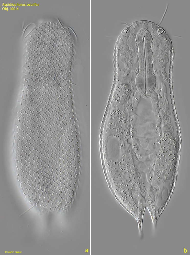
Aspidiophorus oculifer differs from the similar species Aspidiophorus polonicus mainly by the ocelli and the ventral scales. In Aspidiophorus polonicus, the ventral scales are absent or sparsely scattered, and only two narrow, keeled terminal scales are present. Additionally, Aspidiophorus polonicus is somewhat larger, measuring 152–178 µm in length, compared to Aspidiophorus oculifer.
Aspidiophorus-oculifer

Fig. 1 a-b: Aspidiophorus oculifer. L = 100 µm. Two focal planes of a slightly squashed specimen from dorsal. Obj. 100 X.

Aspidiophorus-oculifer

Fig. 2: Aspidiophorus oculifer. A crop from fig. 1 a with details of the head. Apically the ocellar granules (OG) visible. The pharynx (PH) is slightly dumbbel-shaped. Obj. 100 X.

Aspidiophorus-oculifer

Fig. 3: Aspidiophorus oculifer. The dorsal scales in a squashed specimen. Obj. 100 X.

Aspidiophorus-oculifer

Fig. 4: Aspidiophorus oculifer. The detached leave-shaped scales are 3.6–3.9 µm long and a very delicate middle rib is visible. Obj. 100 X.

Aspidiophorus-oculifer

Fig. 5: Aspidiophorus oculifer. L = 111 µm. A squashed specimen from ventral. The two spined terminal scales (TS) are visible. Obj. 100 X.

Aspidiophorus-oculifer

Fig. 6: Aspidiophorus oculifer. The small keeled ventral scales (KS) become only visible in a strongly squashed specimen. TS = terminal scales. Obj. 100 X.