body shield-shaped, posterior margin truncate or slightly convex
dosal side convex with distinct spine
length 35–50 µm, width 30–40 µm
macronucleus horseshoe-shaped
one globular micronucleus adjacent to macronucleus
contractile vacuole on right side, posterior half
7 ventral cirri
5 transverse cirri
frontal membrane of 3 cirri (hard to see)
oral apparatus left, posterior half
adoral zone of 12 membranelles
Aspidisca turrita
I find Aspidisca turrita only very rarely and so far only in the Simmelried and the Ulmisried. Seen dorsally or laterally, the species can be quickly and reliably identified by the conspicuous spine on the convex dorsal side (s. figs 2 a-c and 3 a-c). From a ventral view, identification is more difficult because this species also has the typical Aspidisca ciliature of 7 ventral cirri and 5 transveral cirri (s. fig. 1 a-c). However, the outline is shield-shaped or like an upside-down U. The posterior margin of the body is usually only slightly convex or even straight.
To examine the lateral view with the conspicuous spine, it has proved useful to add a few detritus flakes and the specimens found under a coverslip with hight layer thickness. The specimens quickly find the detritus flakes and begin to run around on them. With increasing evaporation and reduced layer thickness, the specimens can only run around on the lateral sides of the detritus flakes and are then easy to observe.
Aspidisca turrita can be distinguished from the similar species Aspidisca lynceus (smooth dorsal side) and Aspidisca cicada (ribbed dorsal side) by the dorsal spine.
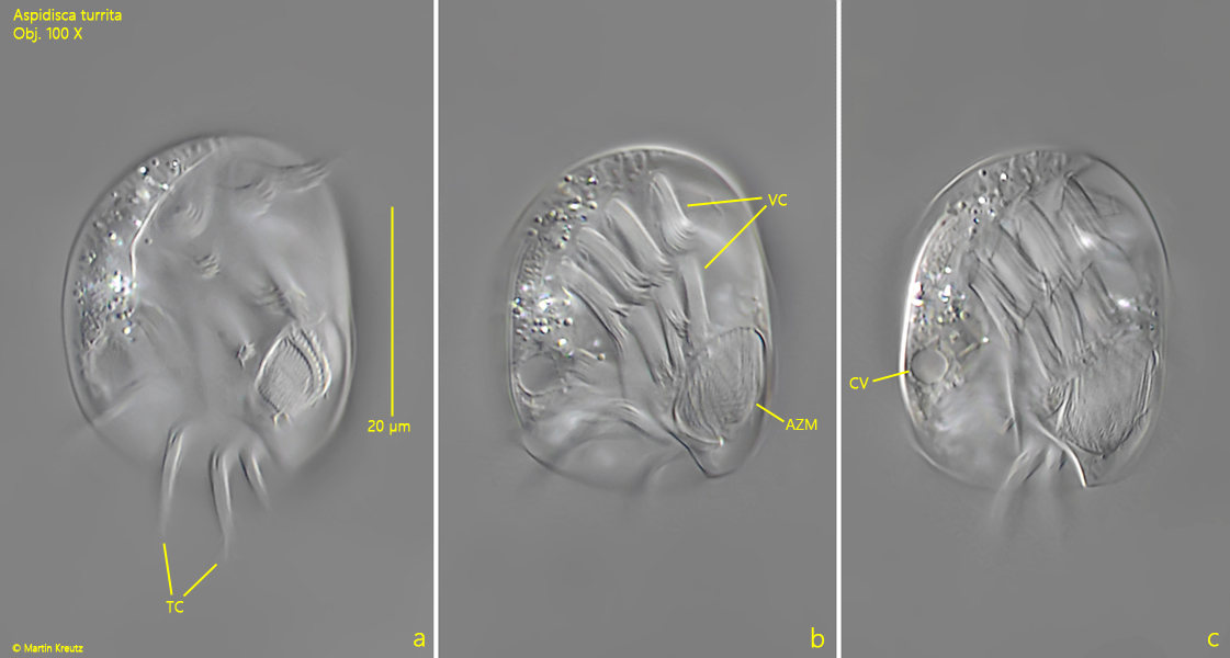
Fig. 1 a-c:Aspidisca turrita. L = 34 µm. Three focal planes of a specimen from ventral. Note the membranelles of the adoral zone (AZM) in the oral apparatus. TC = transverse cirri, VC = ventral cirri. Obj. 100 X.
Fig. 2 a-c:Aspidisca turrita. µm. Three focal planes of the dorsal side with the distinct spine (DS). The spine is laterally flattened, similar to a dorsal fin. Obj. 60 X. Obj. 60 X.
Fig. 3 a-c:Aspidisca turrita. L = 47 µm. A specimen crawling over a detritus flake in lateral view from left. Note the distinct dorsal spine (DS) and the small frontal membranelle (FM) consisting of three cirri. TC = transverse cirri. Obj. 60 X.