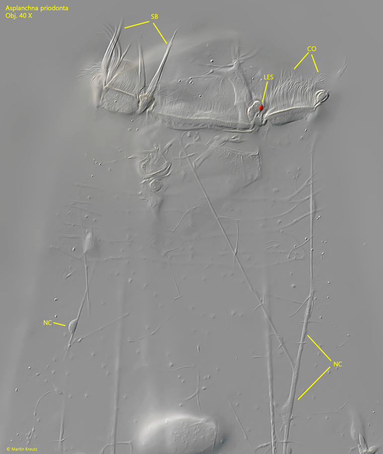
Asplanchna priodonta is one of the most common planktonic rotifers and can be found in almost every lake or pond. Additionally, the species appears to be very adaptable regarding water quality.
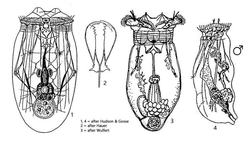
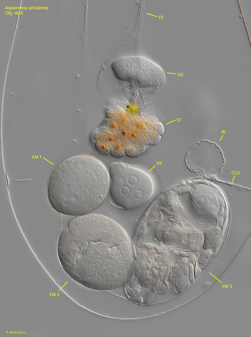
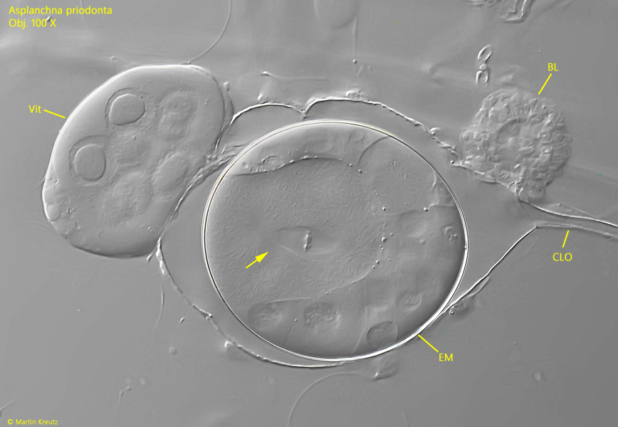
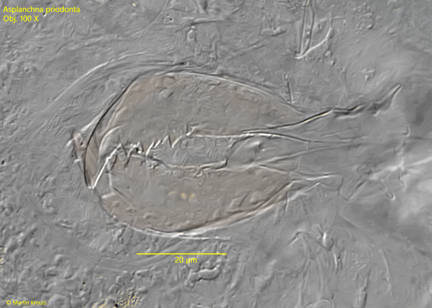
For the identification of Asplanchna priodonta, the shape of the vitellarium and the precise shape of the pincer-shaped rami are important. The vitellarium is spherical and contains very large nuclei with a central nucleolus (s. figs. 4 and 5). The rami are very large, about 50 µm in length, and have distinct teeth on the inner margin (s. fig. 8). They serve to hold the prey and pull it into the pharynx. The similar species Asplanchna girodi has a horseshoe-shaped vitellarium, and the rami lack teeth on the inner side.
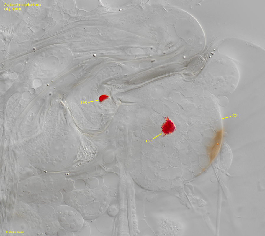
Due to its size and abundance, Asplanchna priodonta is very suitable for the study of the internal structure of rotifers. The organs in the body cavity are clearly separated from each other and can thus be more easily identified (s. fig. 4). In contrast to many other rotifers, however, Asplanchna priodonta lacks an intestine. Therefore, food digested in the stomach is expelled again through the esophagus and mouth opening, as there is no anus.
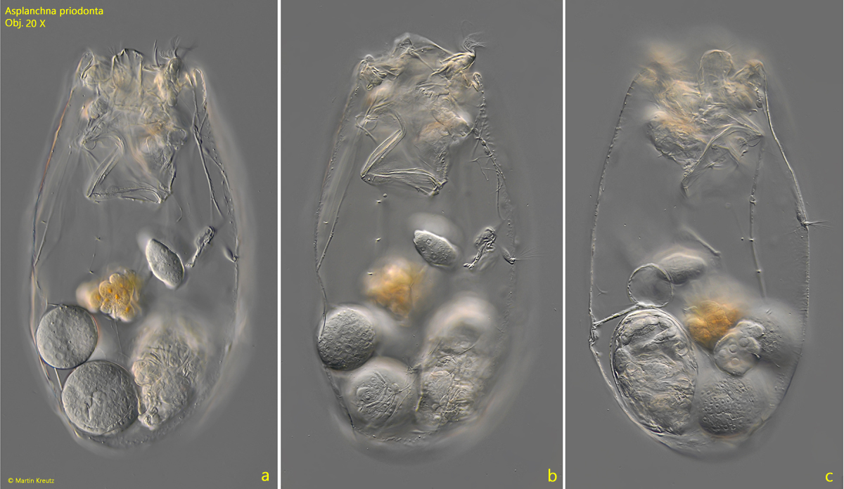
Asplanchna priodonta has a high reproduction rate, allowing habitats to be quickly colonized and occupied. In asexual reproduction (parthenogenesis), unfertilized eggs from the ovary (hardly visible, attached to vitellarium) are supplied with nutrients by the vitellarium and released into the oviduct. There, the development into an embryo takes place through cell division and differentiation. In Asplanchna priodonta, several embryos are usually found in the oviduct, exhibiting different developmental stages (s. fig. 4). The closer they are to the vitellarium, the earlier and less mature the stage. Thus, embryonic development can be tracked like a string of pearls.
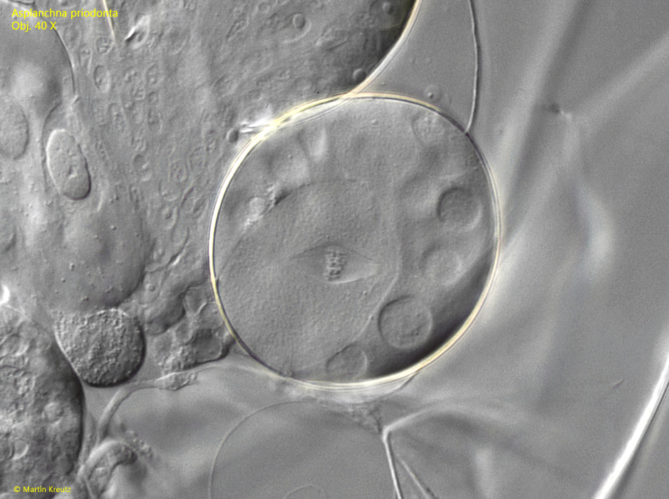
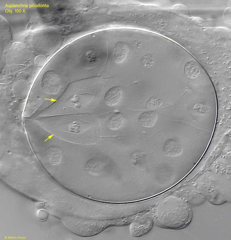
In some embryos at an early, few-celled developmental stage, I was able to observe condensed chromosomes that were about to be separated by the spindle apparatus (s. figs. 5. 6 and 7). In all cases, the cells in which this process occurred were very large. Whether this represents a meiotic reduction division to form haploid eggs during embryonic development or a mitotic cell division, I could not determine.
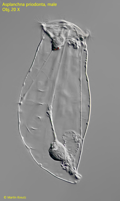
In rare cases, male specimens of Asplanchna priodonta are also found (s. fig. 9). These are about half the size of the female specimens and possess a greatly reduced set of organs. The entire digestive system is absent, and essentially only the seminal vesicle, penis, and urinary bladder are present. The male specimens do not consume any food during their lifetime.
More images and information on Asplanchna priodonta: Michael Plewka-Freshwater life-Asplanchna priodonta