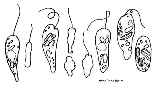
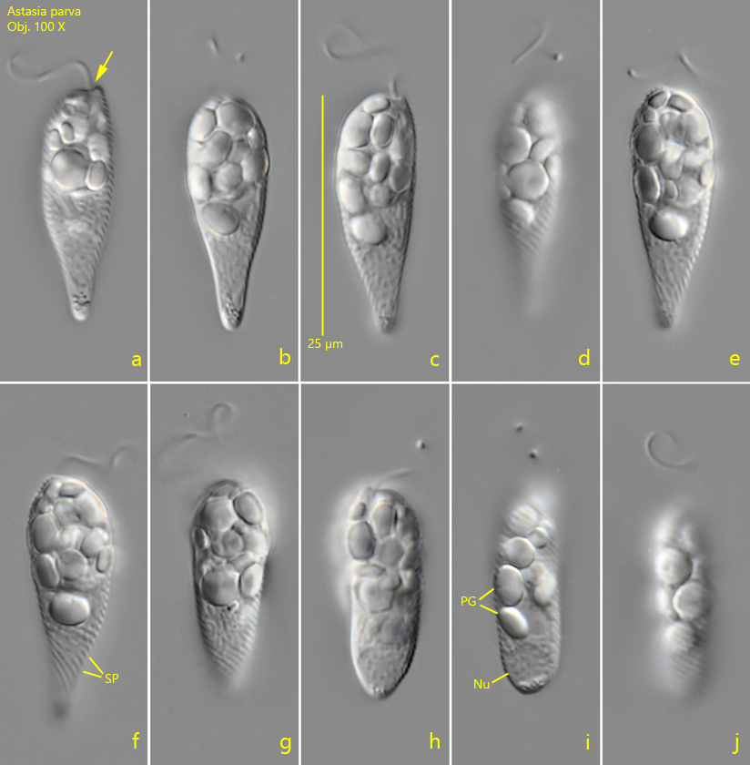
I found several specimens of Astasia parva in the Simmelried in April 2026. All specimens came from the upper layer of mud. This small species was first described by Pringsheim in 1942. It is characterized by a comparatively constant cell shape, which is essentially club-shaped (s. fig. 1 a-e). During swimming, this shape is only slightly altered. Only rarely does a metabolic transition to a more cylindrical form occur (s. fig. 1 g-j). The specimens in my population were all about 25 µm long, thus falling within the range given by Pringsheim.
The only deviation from Pringsheim’s description of Astasia parva was that the paramylon grains in my population were rather round, ellipsoid, and often somewhat flattened (s. fig. 1 i). They were by no means rod-shaped. Nevertheless, I believe that this is Astasia parva, since the alternatives Astasia clava and Astasia gomphonema have different characteristics. Both form head-like widenings at the anterior end, which I was never able to observe in my population. In addition, Astasia clava is plumper and is said to be highly metabolic.
Fig. 1 a-j:Astasia parva. L = 25 µm. A freely swimming specimen. Note that the flagellum arise laterally shifted in a depression (arrow). Nu = nucleus. PG = paramylon grains, SP = counterclockwise running striation of the pellicle. Obj. 100 X.