perizonal stripe of 5 ciliary rows, long as adoral zone
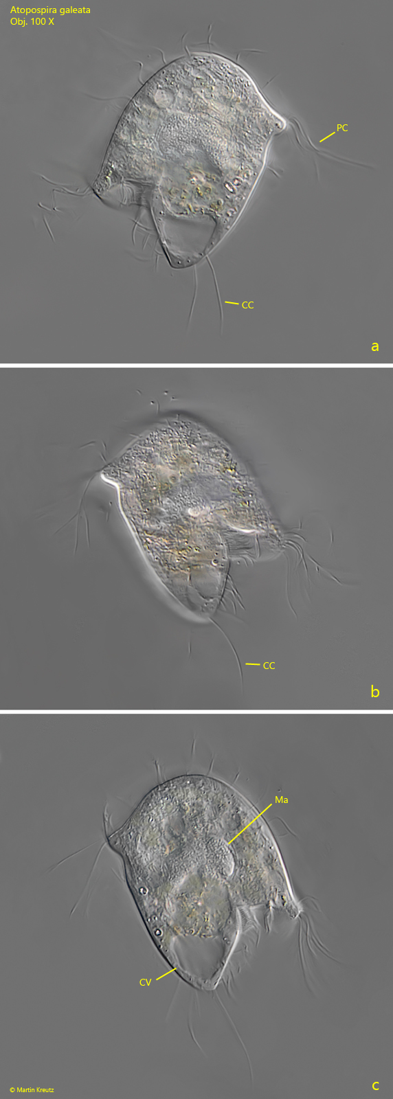
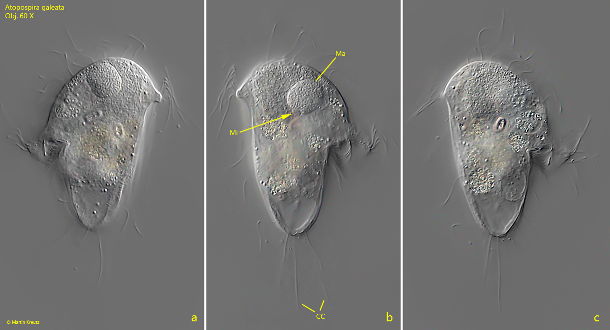
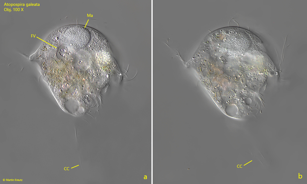
macronucleus globular or broadly oval, located in apical dome
spherical micronucleus adjacent to the macronucleus
cytoplasm filled with ingested sulfur- and rhodobacteria
contractile vacuole terminal
posterior end with long caudal cilia
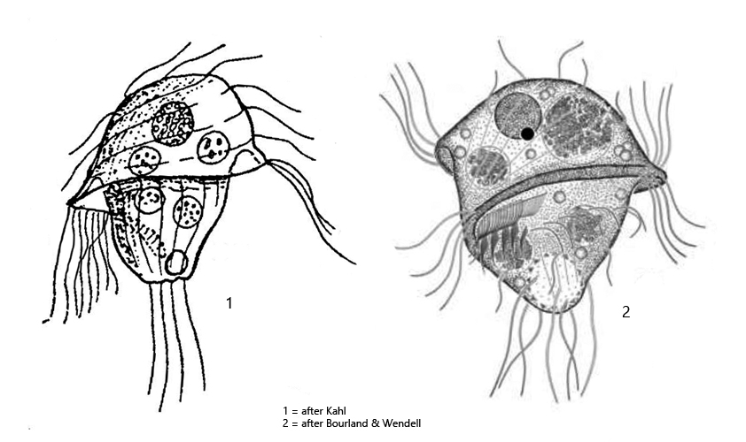
Atopospira galeata
Atopospira galeata was first described in 1927 by Kahl as Metopus galeatus. In 2014 the species was redescribed by Bourland & Wendell who transferred the species to the genus Atopospira and renamed in Atopospira galeata. The genus Atopospira was erected by Jankowski in 1964.
So far I could find Atopospira galeata only in the Simmelried, where the species occurs very rarely. The specimens shown below were found in January 2008, February 2019 and August 2021.
Even at low magnification Atopospira galeata is easily recognized by its helmet-shaped apical dome, which is slightly upturned at the margins (s. fig. 1 a). The species is coverslip sensitive, which is why I could only take images of freely swimming specimens. Most specimens are filled with food vacuoles full of sulfur and rhodobacteria, making them appear yellowish-green or pink in color. The margin of the apical dome bears a wreath of very long cilia, which perform a ruderate movement (s fig. 4 a). The long caudal cilia are delicate and are quickly shed.
Fig. 1 a-c:Atopospira galeata. L = 54 µm. A freely swimming specimen from ventral (a) and from dorsal (b, c). CC = caudal cilia, CV = contractile vacuole, Ma = macronucleus, PC = perizonal cilia. Obj. 100 X.
Fig. 2 a-c:Atopospira galeata. L = 66 µm. A second freely swimming specimen from ventral (a) and from dorsal (b, c). CC = caudal cilia, Ma = macronucleus, Mi = micronucleus. Obj. 60 X.
Fig. 3 a-b:Atopospira galeata. L = 61 µm. A third freely swimming specimen from dorsal. CC = caudal cilia, FV = food vacuoles with ingested sulfur- and rhodobacteria, Ma = macronucleus. Obj. 100 X.
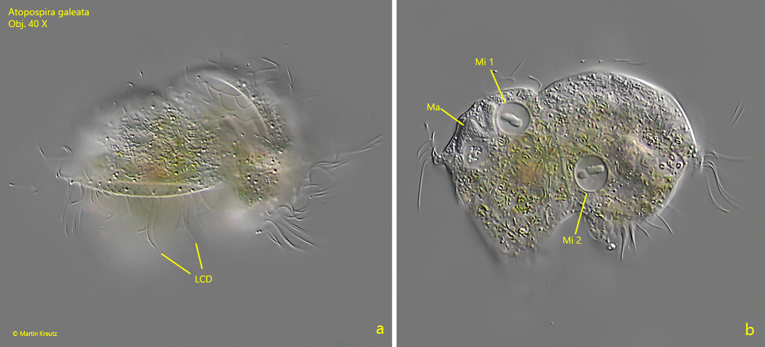
Fig. 4 a-b:Atopospira galeata. Two specimens in conjugation. Note the long cilia on the edge of the helmet-shaped dome (LCD) and the condensed chromosomes in the micronuclei of both specimens (Mi 1, Mi 2). Both specimens are 65 µm long. Obj. 40 X.