I found Basichlamys sacculifera in the Simmelried in March 2022 and, two years later, in January 2025 in the pond of the convent Hegne. The specimens accumulated at the surface in the samples.
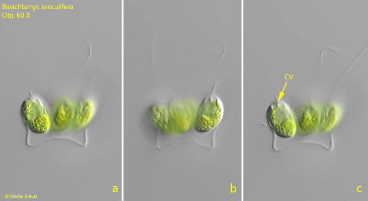
The coenobia are sensitive to a coverslip placed on them and denature quickly. In order to document the natural form I tried to photograph the freely swimming specimens. Although the resolution is greatly reduced at the high layer thickness required for this, all essential features can be seen.
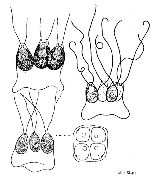
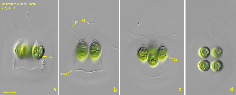
The 4 cells sit in a gelatinous envelope and are held together by it. This envelope consists of the gelatinized cell wall of the mother cell. The concave-shaped base of this envelope is typical. The cells possess two flagella of equal length (s. fig. 2 b) and two contractile vacuoles at the anterior end (s. fig. 1 c). The eyespot is located near the cell equator (s. fig. 2 c). The pyrenoid is located near the posterior end (s. fig. 2 a). The cells themselves were consistently 13–14 µm long and more or less egg-shaped in my population.
Fig. 1 a-c:Basichalmys sacculifera. L = 13 µm (of cells). A freely swimming coenobium in lateral view. Note the two contractile vacuoles (CV) at the anterior end. Obj. 60 X.
Fig. 2 a-d:Basichalmys sacculifera. L = 13–14 µm (of cells). A second freely swimming coenobium. Note the eyespot (ES) in equatorial position. The cells sit in a cub-shaped mucilaginous envelope (CME). Each cell has two flagella (F1, F2) of equal length. Obj. 40 X.