body spindle-shaped, laterally flattened, with tail-shaped posterior end
length 70–165 µm
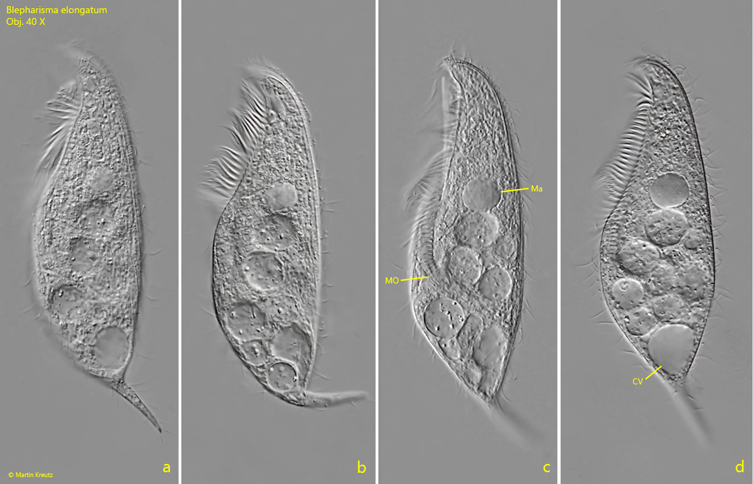
adoral zone on left side of oral groove, with long cilia
undulating membrane inconspicuous, on right side of oral groove
mouth opening below cell equator
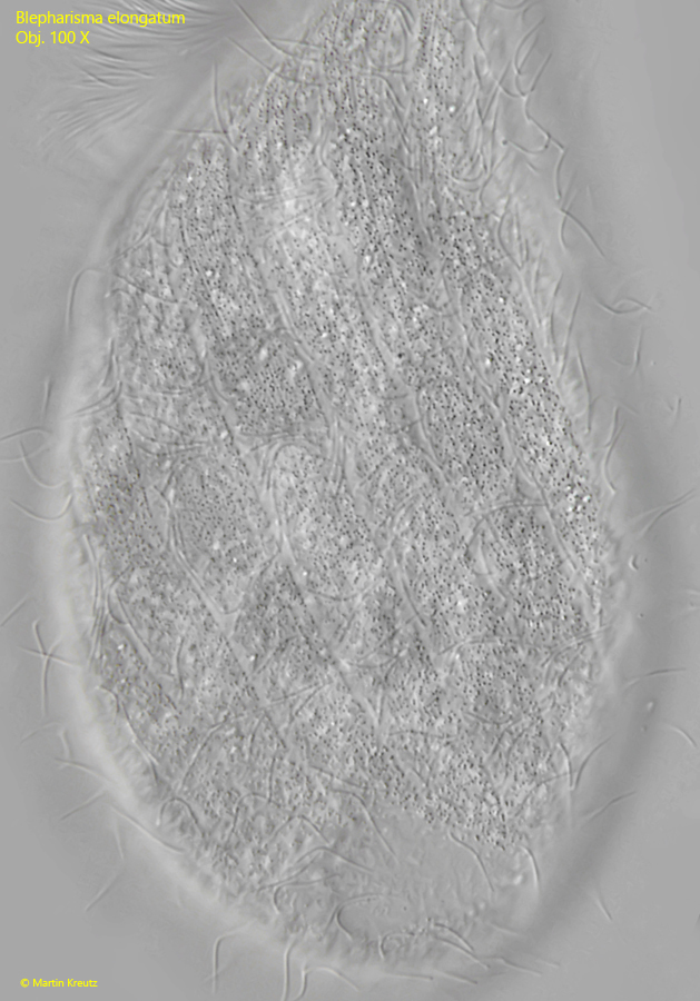
cytoplasm and cortical granules colorless
macronucleus spherical or oval
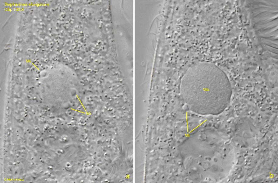
4–7 spherical micronuclei, adjacent to macronucleus
contractile vacuole before base of tail
Blepharisma elongatum
So far, I have found Blepharisma elongatum only once, in June 2025 in an approximately 3‑week‑old sample from the Schwemm Moor in Austria. In the sample I found several specimens, which were located among partially dead algae at the bottom of the vessel.
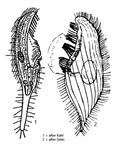
Blepharisma elongatum was first described by Stokes in 1884. Further descriptions were then provided by Kahl (1926) and Foissner (1980). The most recent study is by Larsen & Nilsson from 1988. These authors were also able to maintain Blepharisma elongatum in culture.
The body shape of Blepharisma elongatum resembles that of Blepharisma musculus, as the end of the body is developed into a tail. This is pronounced to varying degrees. The posterior end may also be merely pointed. The essential difference from Blepharisma musculus is the round or oval macronucleus and the colorless granules, which are arranged in broad bands in the pellicle (s. fig. 3). The specimens kept in culture by Larsen & Nilsson showed no signs of coloration even after weeks in darkness.
In Blepharisma elongatum, the macronucleus is surrounded by 4–7 small and round micronuclei. In my population, there were 4–5 micronuclei (s. fig. 2 a-b). Foissner reported in 1980 that he was only ever able to identify a single micronucleus in the two populations he examined. In the cultures of Larsen & Nilsson, however, there were always several micronuclei present.
Fig. 1 a-d:Blepharisma elongatum. L = 164 µm. A freely swimming specimen from left. Note the spherical macronucleus (Ma). The mouth opening (MO) is shortly below the cell equator. CV = contractile vacuole. Obj. 40 X.
Fig. 2:Blepharisma elongatum. Two focal planes of the globular macronucleus (Ma) with a least 5 adjacent micronuclei (Mi). Obj. 100 X.
Fig. 3:Blepharisma elongatum. Focal plane on the rows of colorless granules beneath the pellicle. Obj. 100 X.