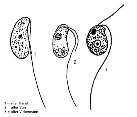
one leading flagellum of body length (hard to see)
one trailing flagellum, 2–3 body length, curved
contractile vacuole subapical
spherical nucleus below cell equator
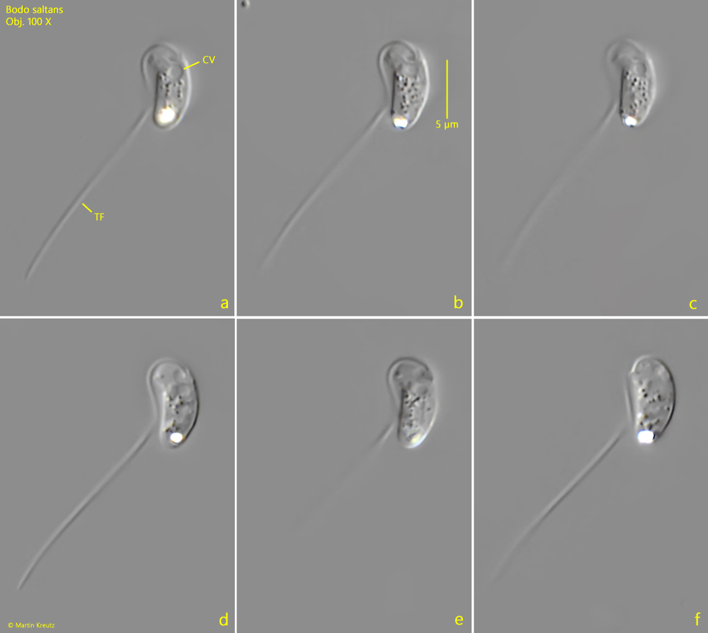
I occasionally find Bodo saltans on the floating coverslip, where it can be easily observed. This small flagellate can mainly be recognized by its long trailing flagellum, which forms a sharp curve and touches the body, while the distal part of the flagellum is held more or less straight and spread away from the body (s. fig. 1 a). It is 2–3 times as long as the body. There is also a second swimming flagellum, but it is difficult to see as it often lies flat against the body in a shallow groove.
Bodo saltans exhibits a conspicuous, jumping-jerking movement. This is caused by the swimming flagellum, which pops out briefly and then lies flat again. Bodo saltans can attach itself to surfaces with the end of its trailing flagellum. Several specimens are often found attached to detritus flakes, but they are then difficult to observe in detail.
Bodo saltans belongs to the euglenids and has only a single mitochondrion, which is tubular in shape. In the apical part of the mitochondrion there is a DNA-rich thickening called the kinetoplast. The kinetoplast has a higher density than the surrounding cytoplasm and can sometimes be seen under a light microscope.
Fig. 1 a-f:Bodo saltans. L = 7.7 µm. Different focal planes of a freely swimming specimen. The trailing flagellum (TF) is strongly curved and touches the body. The shorter swimming flagellum is not visible, as it often lies flat against the body in a shallow groove. CV = contractile vacuole. Obj. 100 X.