So far, I have found Brachionus diversicornis only twice. The first find was in June 2006 in Mühlhalden Pond. It was not until 17 years later, in August, that I found a second population in the pond at the Fliesshorn campsite.
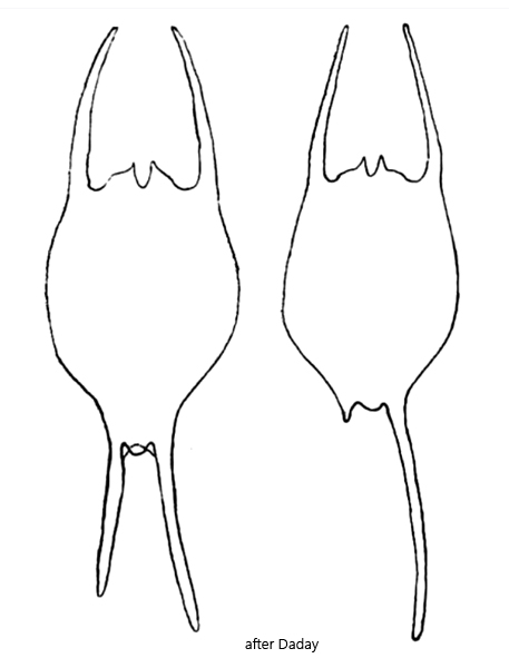
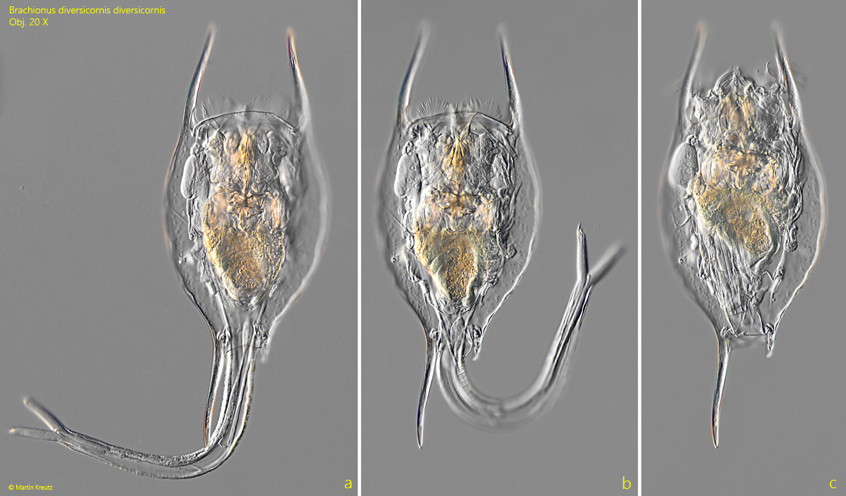
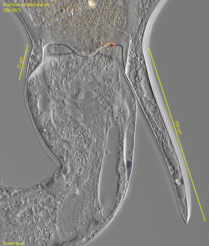
Brachionus diversicornis is very prominent in plankton samples because of its size. The specimens can also be recognized very easily by the shape of their lorica, and especially by the two posterior spines of different lengths. Remarkably, the right spine is almost always longer than the left one (s. fig. 5).
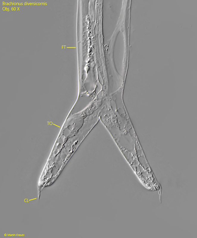
The long, ringed foot is only ever extended very briefly. It then lashes back and forth like a whip and is immediately retracted again (s. fig. 1 a and 1 b). At the end, the foot splits into two finger-shaped toes. These in turn bear short, very pointed claws (s. fig. 6). Sometimes these can also occur in pairs.
Brachionus diversicornis has already been documented on all continents except North America. The reasons for this are unknown.
Fig. 1 a-c:Brachionus diversicornis. L = 420 µm (with spines). A slightly squashed specimen from ventral. Note the longer right posterior spine compared to the left spine. Obj. 20 X.
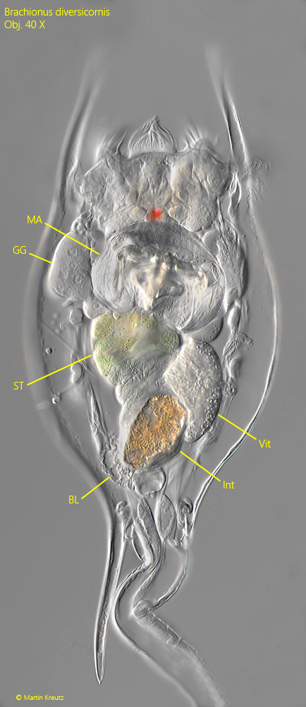
Fig. 2:Brachionus diversicornis. L = 430 µm (with spines). A second slightly squashed specimen from ventral at higher magnification. BL = bladder, GG = gastric gland, Int = intestinum, MA = mastax, ST = stomach, Vit = vitellarium. Obj. 40 X.
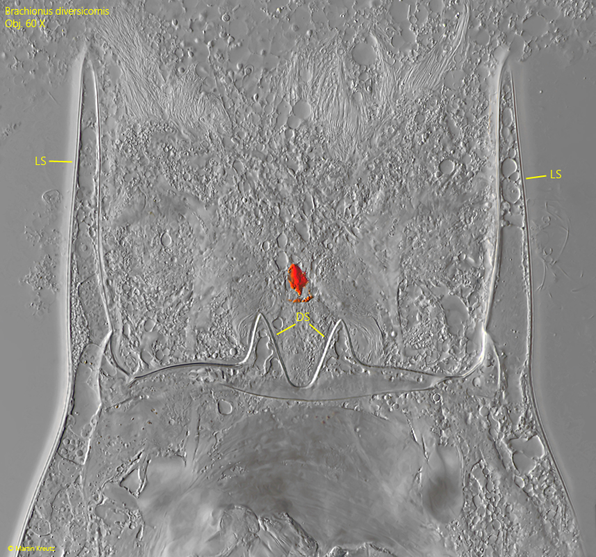
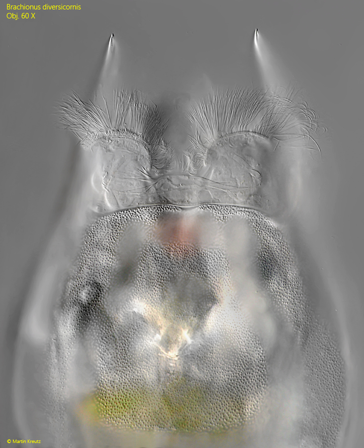
Fig. 3:Brachionus diversicornis. The two dorsal anterior spines in a strongly squashed specimen. LS = lateral anterior spines. Obj. 60 X.
Fig. 4:Brachionus diversicornis. The ventral anterior marging with a slight indentation in the middle. The lorica is finely granulated. Obj. 20 X.
Fig. 5:Brachionus diversicornis. The two posterior spines in a strongly squashed specimen from dorsal. The right spine is 108 µm long while the left spine is only 21 µm long. Obj. 60 X.
Fig. 6:Brachionus diversicornis. The squashed foot (FO) with two toes (TO). At the end of the toes pointed claws (CL) are visible. Obj. 60 X.