Bryceella perpusilla

Wilts, Arbizu & Ahlrichs, 2010

Most likely ID: n.a.


Synonym: n.a.


Sampling location: Moss


Phylogenetic tree: Proalidae


Diagnosis: 

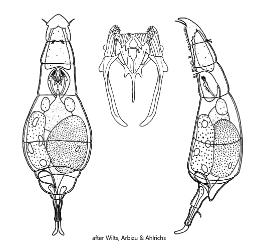
  • body fusiform, dorso-ventrally flattened
  • length 50-80 µm
  • rostrum triangular
  • head consists of 3 pseudosegments
  • trunk consists of 4 pseudosegments
  • corona on ventral side, one fifth of body length
  • head with two lateral styli
  • eyespot absent
  • vitellarium with 4 nuclei
  • foot slender with 2 pseudosegments
  • rounded toes, curved ventrally
  • one caudal antenna
Bryceella-perpusilla
Bryceella perpusilla

I found several specimens of Bryceella perpusilla in March 2017 and in March 2021 in moss samples that I collected from trees. I moistened these in Petri dishes with rainwater. After a few days, the first specimens of Bryceella perpusilla could be found.


Bryceella perpusilla was only described in 2010 by Wilts, Abrizu and Ahlrichs from populations in moss. It is an extremely agile rotifer. Its movements appear hectic and searching. In the process, the specimens constantly contract and stretch, which makes photographing extended specimens difficult.


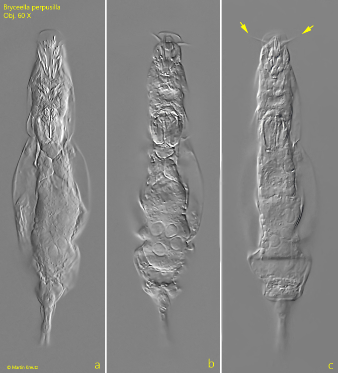
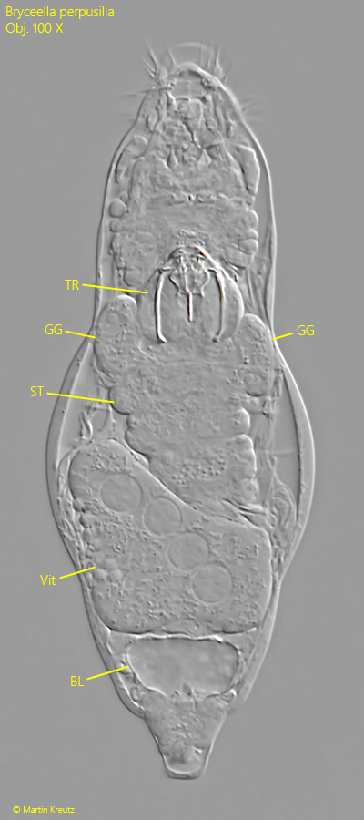
As a characteristic feature, Bryceella perpusilla has two apical, antenna-like styli (s. fig. 2 c). The head has a triangular rostrum that is drawn out ventrally in a cap-like shape. However, this can only be seen in lateral view. The corona consists of several sectors of parallel-arranged cilia (s. figs. 3a and 3d). Only in a squashed specimen can one recognize that the vitellarium contains only 4 nuclei (s. fig. 5). The toes have no claws and are rounded at the end. They are curved toward the ventral side.


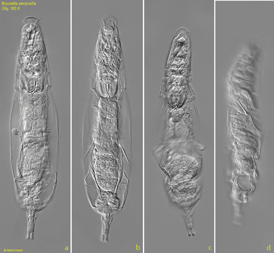
The specimens of my population, with a length of up to 120 µm (extended specimens, s. fig. 4 a-d), were about 30% larger than described by Wilts, Abrizu and Ahlrichs. Since the species has so far only rarely been found and described, little is known about the variability within the genus. All other characteristics agree with the description by Wilts, Abrizu and Ahlrichs.
 

More images and information on Bryceella perpusilla: Michael Plewka-Freshwater life-Bryceella perpusilla

Bryceella-perpusilla

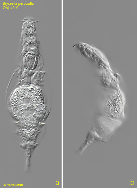
Fig. 1 a-b: Bryceella perpusilla. L = 116 µm. A freely swimming specimen from ventral (a) and from left (b). Obj. 40 X.

Bryceella-perpusilla

Fig. 2 a-c: Bryceella perpusilla. L = 107 µm. A second freely swimming specimen from ventral. Note the two apical styli (arrows). Obj. 60 X.

Bryceella-perpusilla

Fig. 3 a-d: Bryceella perpusilla. L = 107 µm. The same specimen as shown in fig. 2 a-c at higher magnification. Obj. 100 X.

Bryceella-perpusilla

Fig. 4 a-d: Bryceella perpusilla. L = 121 µm. A third freely swimming specimen from ventral (a-c) and slightly contracted from right (d). Obj. 100 X.

Bryceella-perpusilla

Fig. 5: Bryceella perpusilla. A squashed specimen from ventral. Note the 4 nuclei in the vitellarium (Vit). BL = bladder, GG = grastric glands, ST = stomach, TR = trophi. Obj. 100 X.

Bryceella-perpusilla

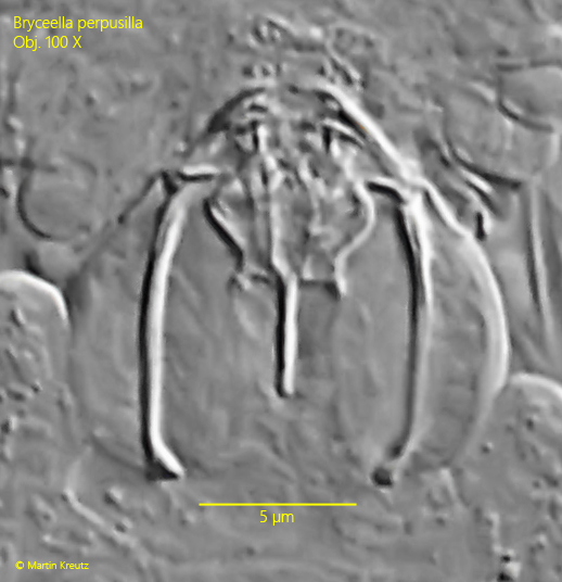
Fig. 6: Bryceella perpusilla. The trophi of the specimen as shown in fig. 5. Obj. 100 X.