Caudiholosticha viridis is a very common hypotrich ciliate, which I find in the upper layer of the sludge zone in the Simmelried and in the Ulmisried. I especially often find the specimens among heavily decomposed leaves. Several times I was able to observe a mass development with over 20 specimens per milliliter.
Berger (2003) transferred all species that possess the characteristics of the genus Holosticha but additionally have caudal cirri into the genus Caudiholosticha. Within the genus Caudiholosticha, only the species Caudiholosticha viridis has symbiotic algae.
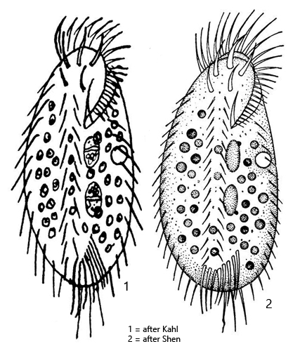
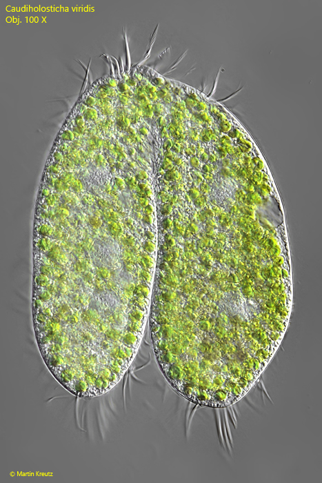
Apparently, there are only a few observations and descriptions of Caudiholosticha viridis. Berger (2003) essentially reproduces the brief description by Kahl (1932). The specimens of my population essentially correspond to Kahl’s description, but my specimens are significantly longer at 130–155 µm and the body shape is slimmer than drawn by Kahl (s. drawing above).
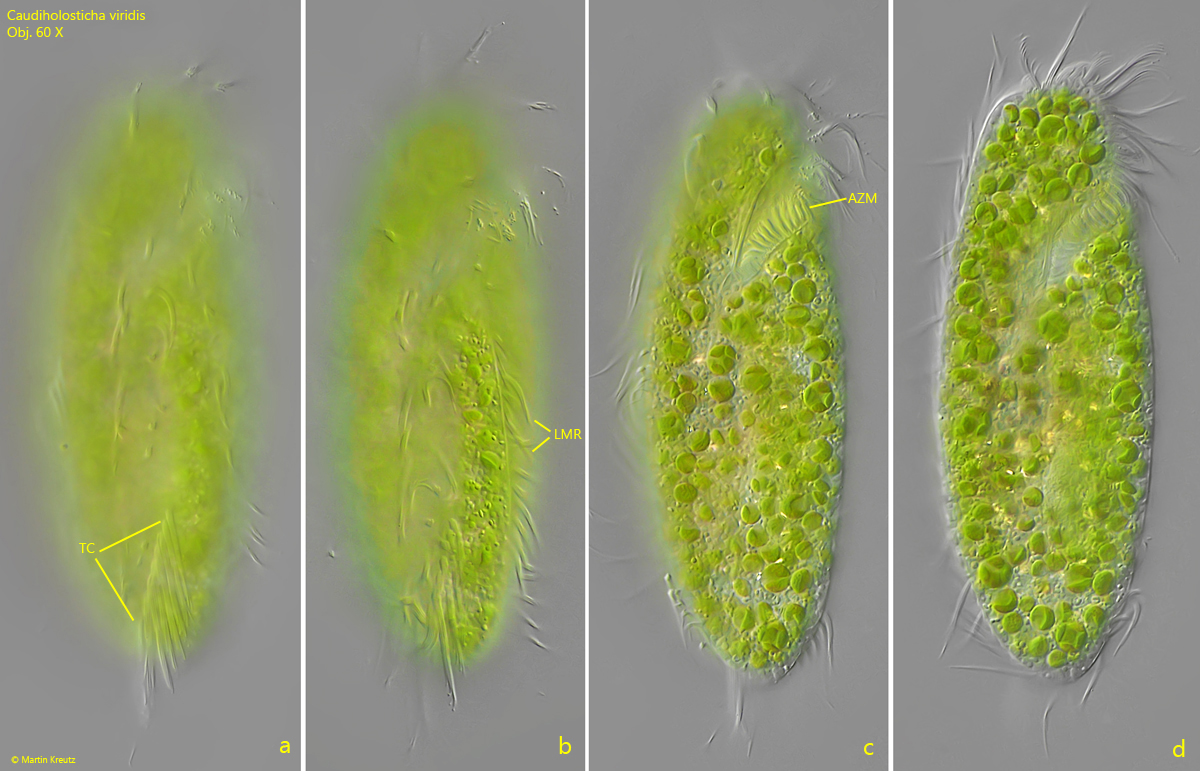
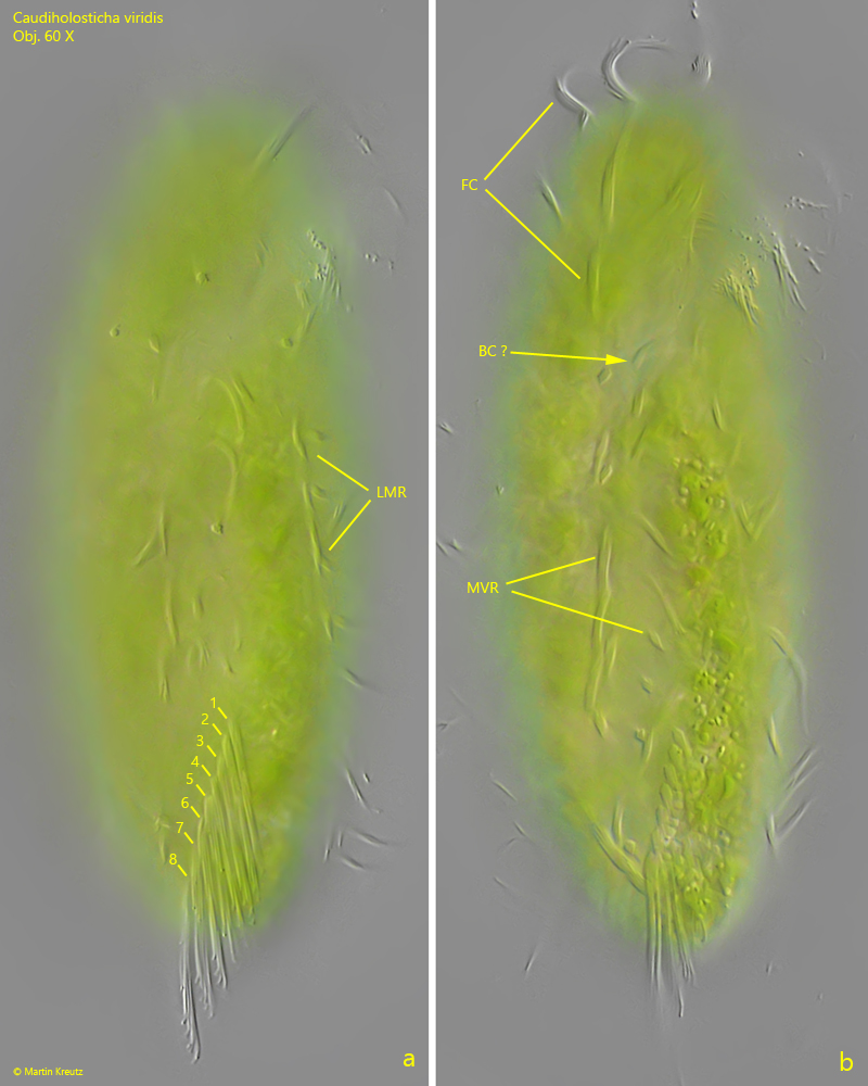
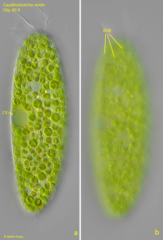
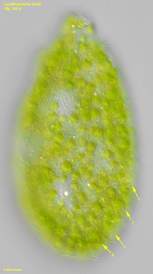
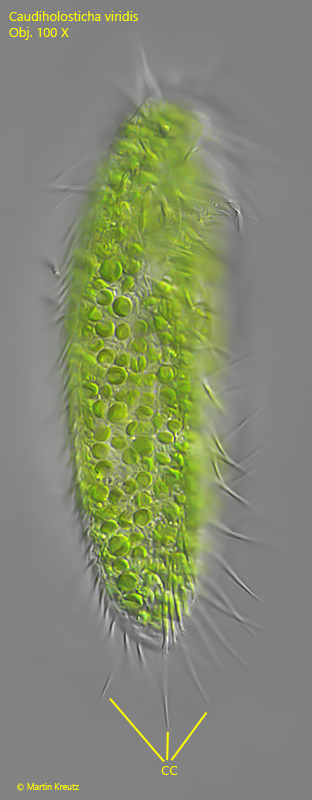
The pattern of the ventral cirri is difficult to discern without staining due to the many symbiotic algae in the cytoplasm. Additionally, the specimens are somewhat sensitive to coverslips and deform quickly. However, I was able to clearly recognize the 8 transverse cirri, which are arranged in a straight, oblique row (s. figs. 1 a and 2 a). There are 2 rows of midventral cirri as well as a right and a left marginal row of cirri (s. fig. 2 a-b). I could not precisely determine the number of frontal cirri and could not clearly identify the buccal cirrus to the left of the mouth opening. The three caudal cirri are clearly visible and represent an extension of the left marginal row of cirri (s. fig. 5).
The dorsal ciliation has not been described so far. According to my observations, there are at least 4 longitudinal rows of bristle-like cilia, which, however, do not run straight across the body. In addition, some of these bristles are arranged in pairs (s. figs. 3 b and 4).
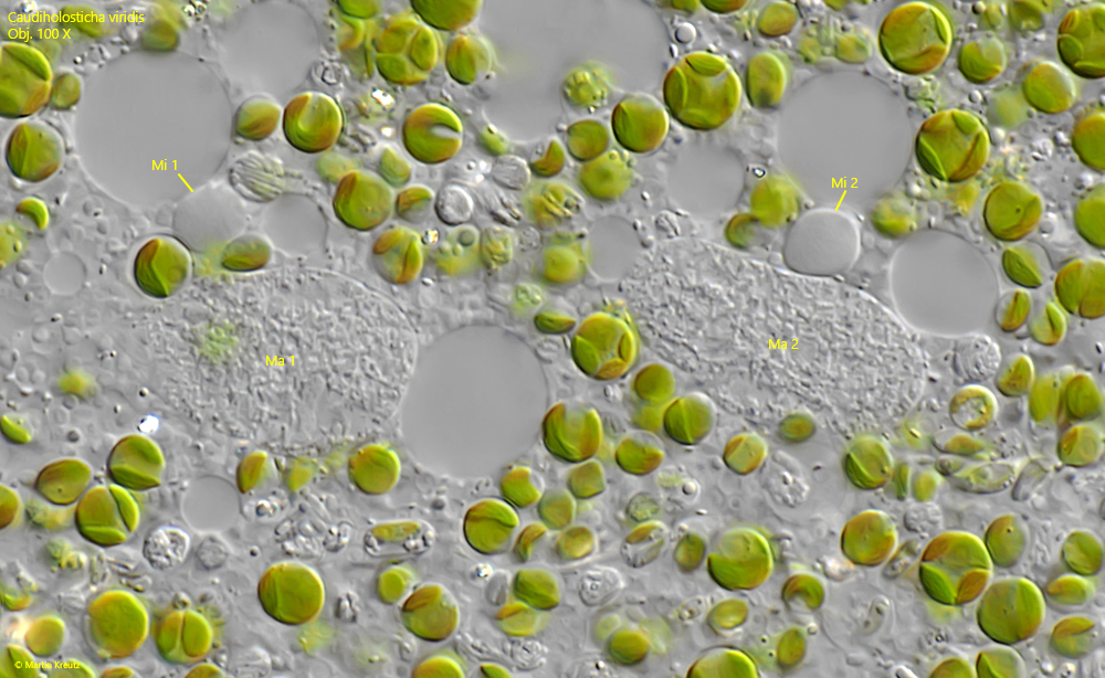
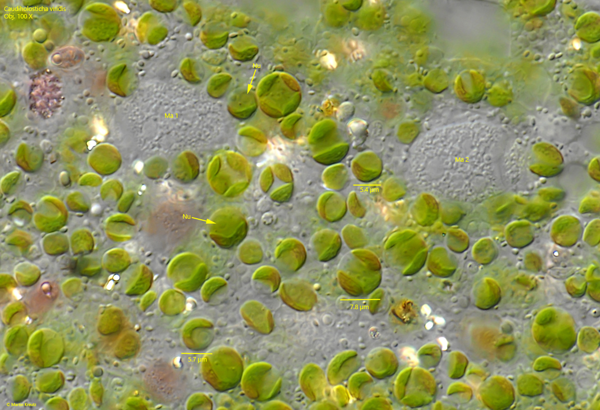
The entire cytoplasm is densely filled with symbiotic algae. These algae were identified by Sud (1969) as Chlorella miniata. According to my observations, these algae have a diameter of 5.4–7.8 µm, no pyrenoid, and their own centrally located nucleus.
The hypotrich ciliates Oxytricha chlorelligera and Paraurostyla viridis also possess symbiotic algae and can be confused with Caudiholosticha viridis. However, these two species have only 5 transverse cirri, no caudal cirri, and no 2 midventral rows of cirri.
In 2021, a publication by Song et al. was released in which Caudiholosticha viridis was subjected to a genetic analysis. As a result, Song et al. transferred Caudiholosticha viridis to the newly established genus Bourlandella (Bourlandella viridis). Since I could not determine whether this taxonomically new classification is valid and recognized, I mention it here but retain the designation Caudiholosticha viridis.