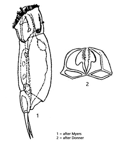
body slender elongated, slightly compressed laterally
length 148–220 µm
corona ventrally oriented, without lips
neck well marked
eyespot absent
ganglion long and sacccate
below ganglion a retrocerebral sac with a distinct duct
lateral cleft of lorica parallel sided
gastric glands large and pigmented orange or reddish
foot conical and short
Cephalodella forficata
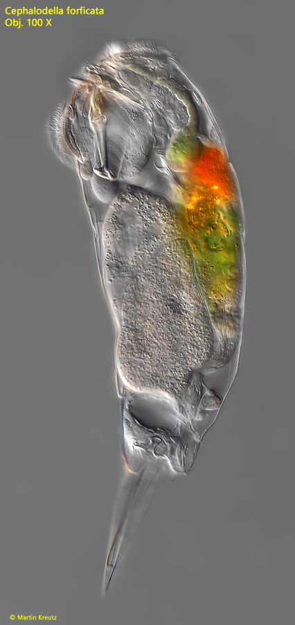
Cephalodella forficata is a common rotifer in my sampling sites. I usually find it between floating plants. The species is easy to recognize even at small magnifications due to the conspicuous orange or reddish coloured gastric glands (s. fig. 2).
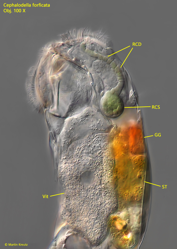
Cephalodella forficata has a retrocerebral sac which is located below the cone-shaped ganglion. Little is known about this organ. At high magnification it can be seen that it is filled with greenish colored, highly refractive vesicles (s. fig. 2). These do not fuse, but remain separate. They are directed to the front end of the head through a retrocerebral duct (s. fig. 2). If a specimen is strongly squashed, oily vesicles are released, which could be the vesicles produced in the retrocerebral sac (s. fig. 4). As the exit of the retrocerebral duct is located in the middle of the corona, it can be assumed that the substances released have something to do with food intake. It may be a kind of “saliva” that is enriched with enzymes and serves to pre-digest food.
Fig. 1:Cephalodella forficata. L = 160 µm. A slightly squashed specimen from left. Obj. 100 X.
Fig. 2:Cephalodella forficata. The head in detail of the specimen as shown in fig. 1. The retrocerebral sac (RCS) is filled with greenish colored vesicles. A retrocerebral duct (RCD) is leading to the apical end of the head. GG = orange colored gastric glands, ST = stomach, Vit = vitellarium. Obj. 100 X.
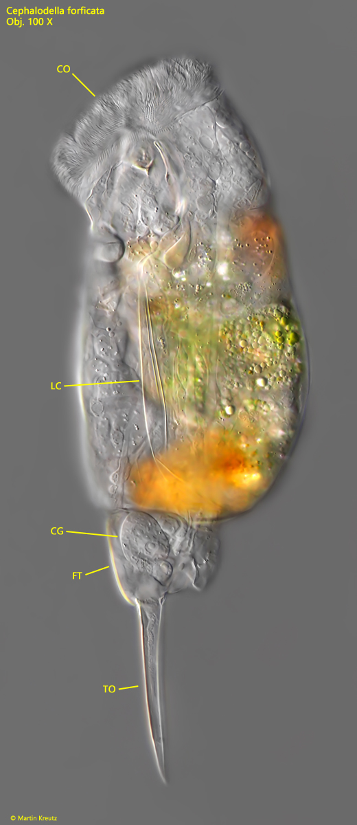
Fig. 3:Cephalodella forficata. L = 192 µm. A stronger squashed specimen from left. The focal plane is on the corona (CO) and the lateral cleft (LC) of the lorica. CG = cement glands, FT = foot, TO = toes. Obj. 100 X.
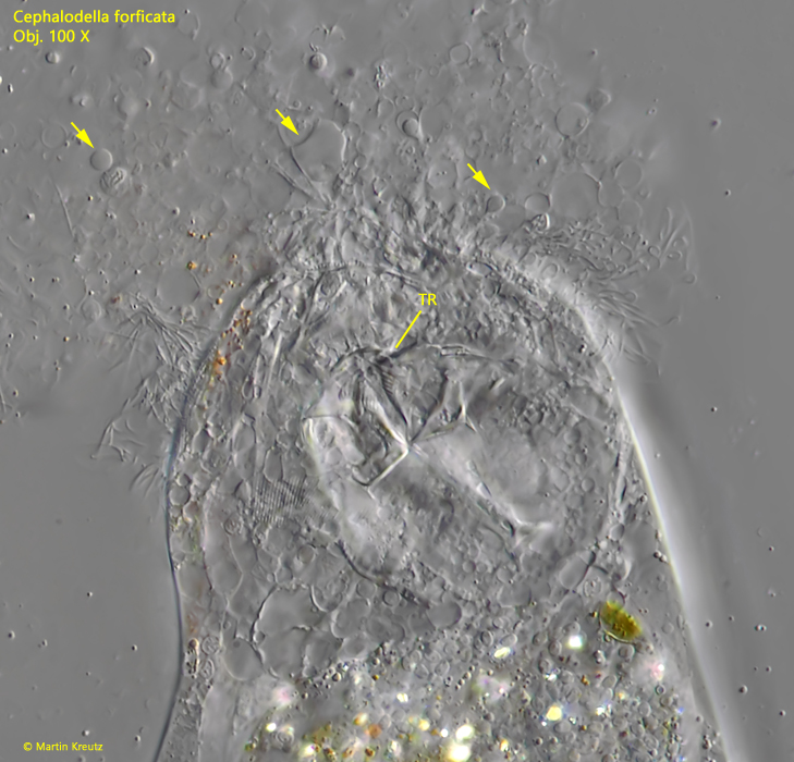
Fig. 4:Cephalodella forficata. In a strongly squashed specimen an oily liquid is released (arrows). It is possible that it is the content of the retrocerebral sac. TR = trophi. Obj. 100 X.