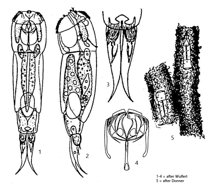
body almost cylindrical, elongated, slightly compressed laterally
length 160–425 µm
corona ventrally oriented
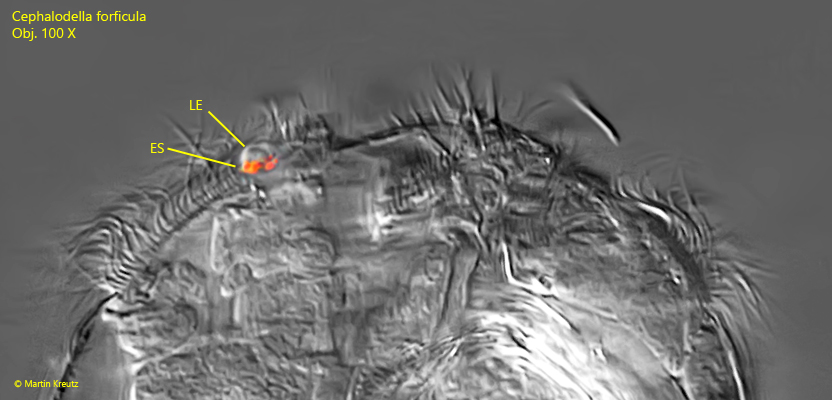
two adjacent eyespots with one lens
toes commonly bent dorsally
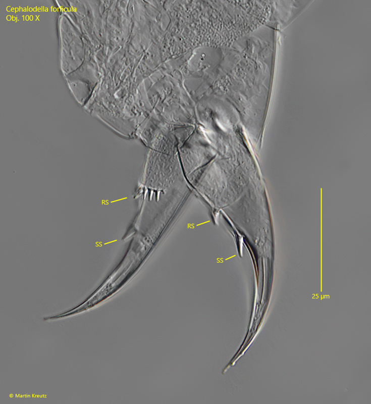
toes with a transverse row of 2–6 short spine and a longer spine below
tube-dwelling species, tubes built up from detritus
Cephalodella forficula
So far I have only found Cephalodella forficula in the Simmelried. The species was very common until 2011. The long, brownish tubes were often found on the walls of the sample jars. I did not find any more specimens after 2011.
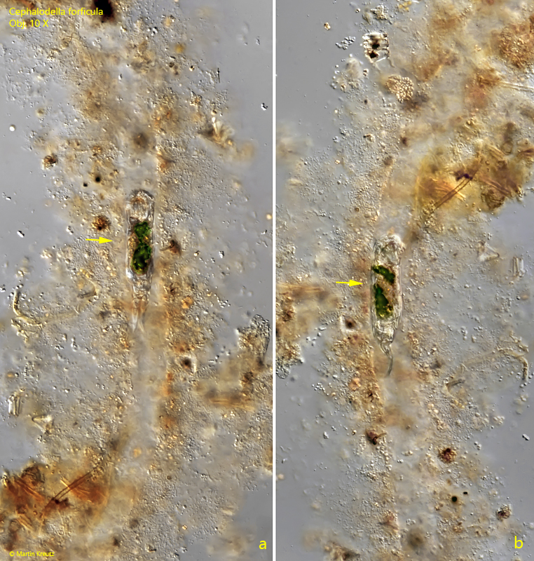
Cephalodella forficula is the only species within the genus Cephalodella that lives in self-constructed tubes. These tubes are built from detritus. The parallel sided tubes have a diameter of 60–70 µm and covered with a thick layer of detritus (s. fig. 6 a-b). Donner (1966) gives the diameter of the tubes as 80 µm.
When disturbed, the specimens leave the tubes and can be observed swimming freely. The specimens are easily recognized by the thick, dorsally curved toes, which bear conspicuous spines (s. fig. 4) and by the apical eyespot with lens (s. fig. 3). The spines on the dorsal side of the toe are assembled as a transverse row of 2–6 short spines and a single longer spine below. The transverse row always consisted of 4 spines in the specimens of my population. The eyespot appears solitary in lateral view (s fig. 3), but it is two eyespots lying close together below a common lens. However, this arrangement can only be recognized in apical view.
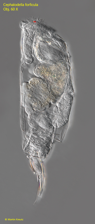
Fig. 1: Cephalodella forficula. L = 240 µm. A freely swimming specimen from right. Obj. 60 X.
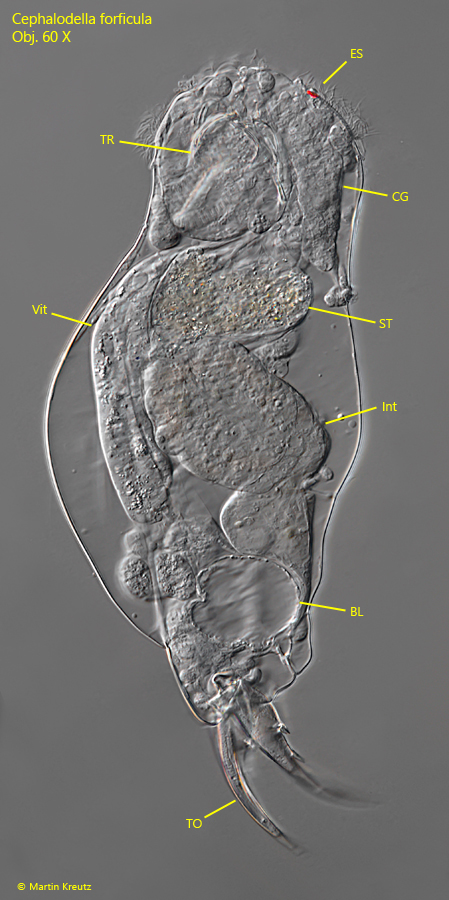
Fig. 2: Cephalodella forficula. L = 240 µm. The slightly squashed specimen as shown in fig. 1 from left. BL = bladder, CG = central ganglion, ES = eyespont with lens, Int = intestine, ST = stomach, TO = toes, Vit = vitellarium. Obj. 60 X.
Fig. 3: Cephalodella forficula. The eyespot (ES) with lens (LE) in detail. The eyespot is biparted, what is only visible in apical view. Obj. 100 X.
Fig. 4: Cephalodella forficula. The dorsally bent toes of a squashed specimen in detail. The toes of this specimen are 56 µm long with a transverse row of several short spines (SS) and a longer single spine (SS) below. Obj. 100 X.
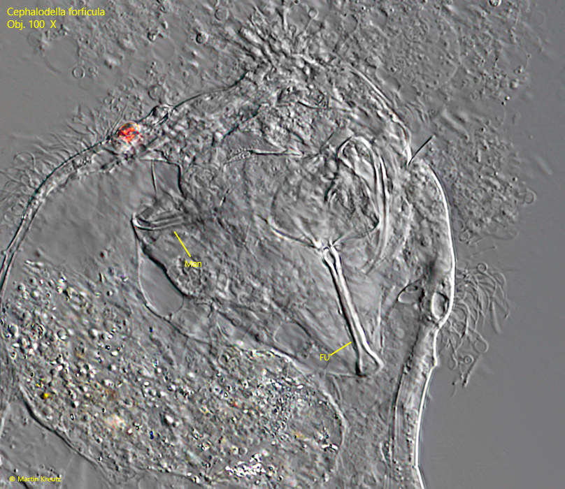
Fig. 5: Cephalodella forficula. The trophi in a squashed specimen. FU = fulcrum, Man = manubrium. Obj. 100 X.
Fig. 6 a-b: Cephalodella forficula. A specimen (arrows) in a self-constructed tube constructed of detritus from dorsal (a) and from right (b). The diameter of the tube is 60–65 µm. Obj. 10 X.