lateral clefts widening gradually to posterior end
foot small, conical
toes long, slightly recurved with conical tips
Cephalodella gibba
Cephalodella gibba is one of the most common species within the genus. I find the species in various locations, mostly among floating aquatic plants.
The specimens of my population were about 250–350 µm long, sometimes a bit smaller. According to Koste (1978), however, there are also giant forms with a length of up to 450 µm. The most striking features of Cephalodella gibba are the distinct and large frontal eyespot and the long, slightly backward-curved toes, which end in a small point (s. fig. 4). The specimens are often yellowish or orange in color. Cephalodella gibba is an omnivore and lives predatory on flagellates, ciliates, and other rotifers, but also feeds on unicellular algae.
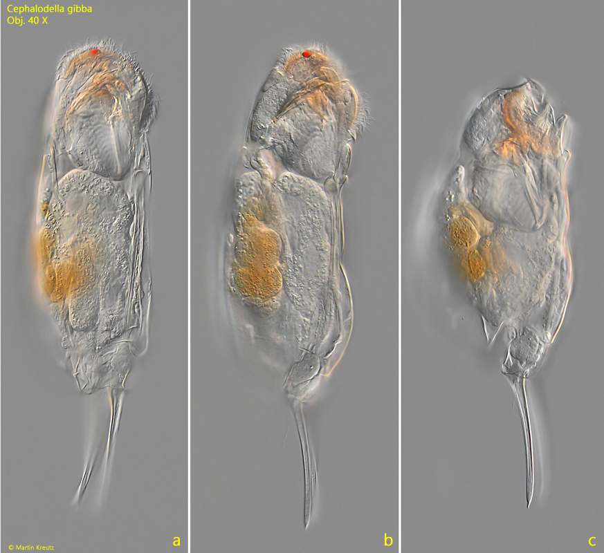
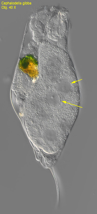
Fig. 1 a-c:Cephalodella gibba. L = 240 µm (with toes). A freely swimming specimen from right. Obj. 40 X.
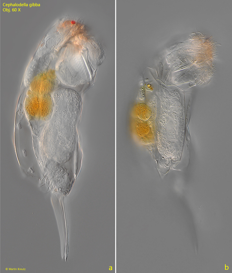
Fig. 2 a-b:Cephalodella gibba. L = 240 µm (with toes). The slightly squashed specimen as shown in fig. 1 a-b. Obj. 60 X.
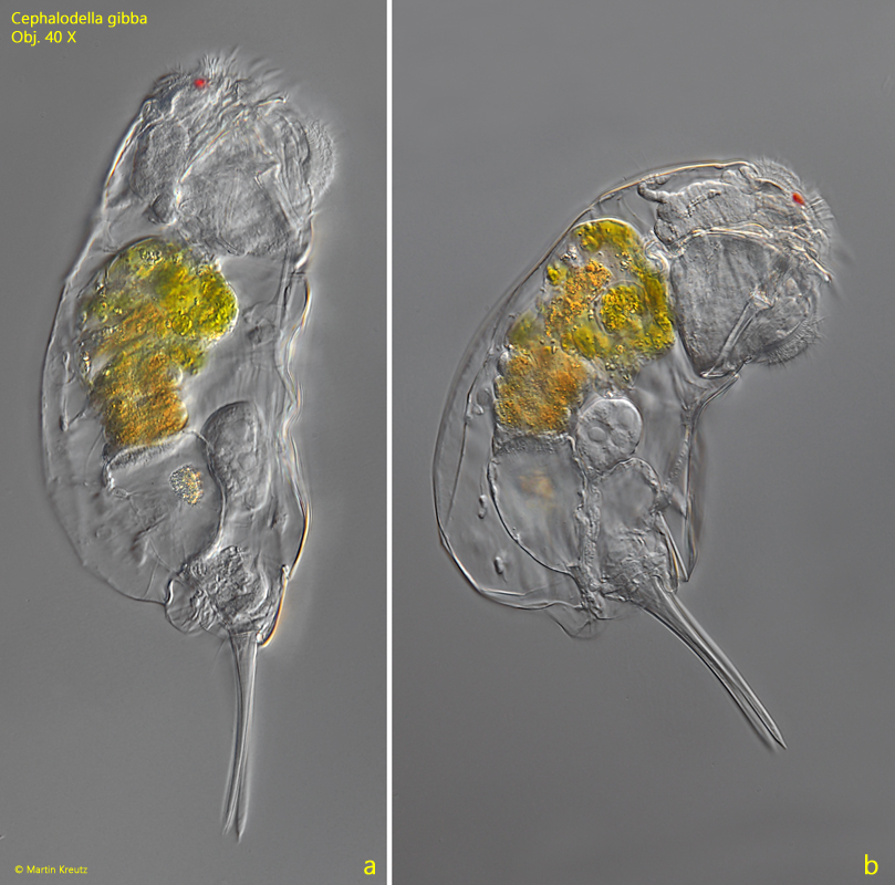
Fig. 3 a-b:Cephalodella gibba. L = 336 µm (with toes). A second specimen found in November 2016 in the Ulmisried. Obj. 40 X.
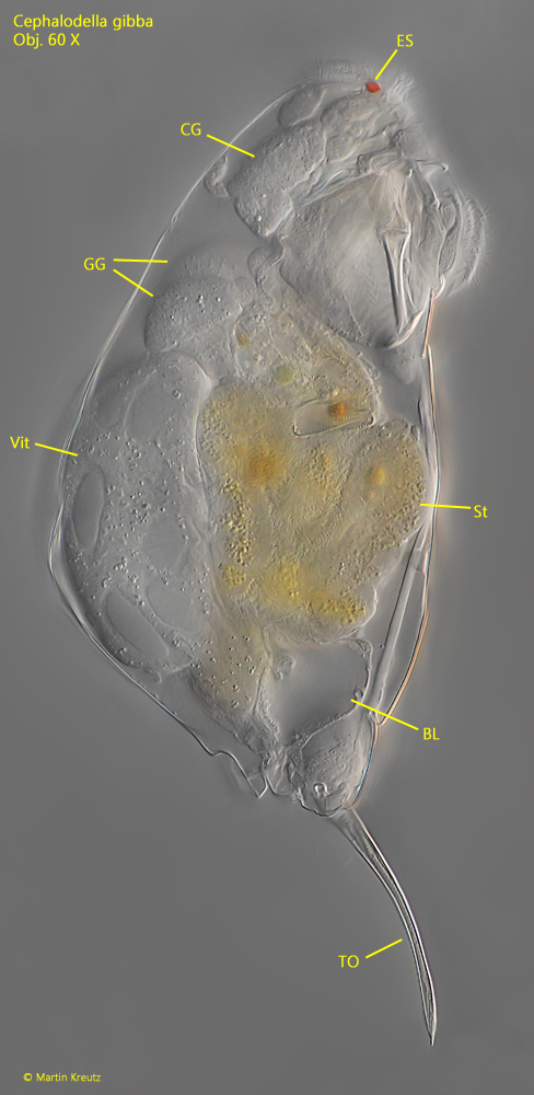
Fig. 4:Cephalodella gibba. L = 275 µm (with toes). The second, slightly squashed specimen. BL = bladder, CG = cerebral ganglion, ES = exespot, GG = gastric glands, St = stomach, TO = toes, Vit = vitellarium. Obj. 60 X.
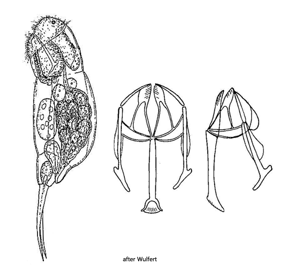
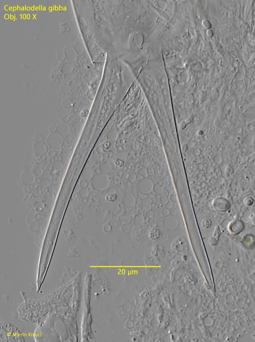
Fig. 5:Cephalodella gibba. The toes with a length of 68 µm in a squashed specimen. Obj. 100 X.
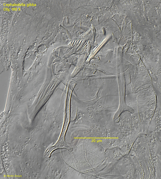
Fig. 6:Cephalodella gibba. The trophi in a strongly squashed specimen. Obj. 100 X.
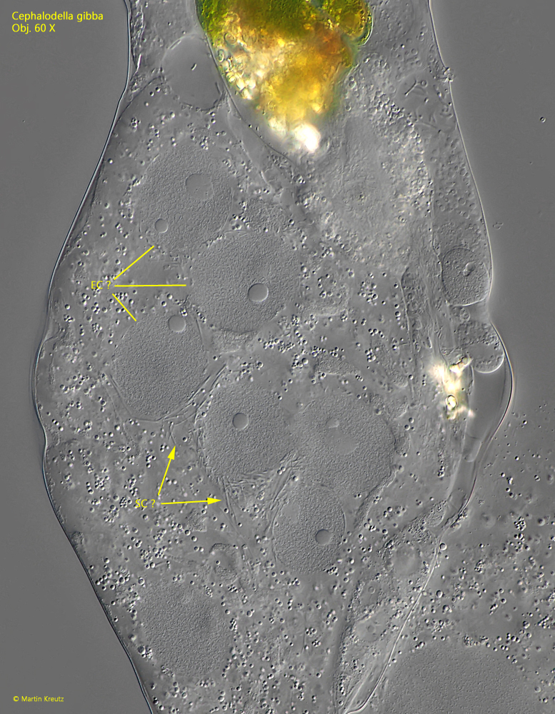
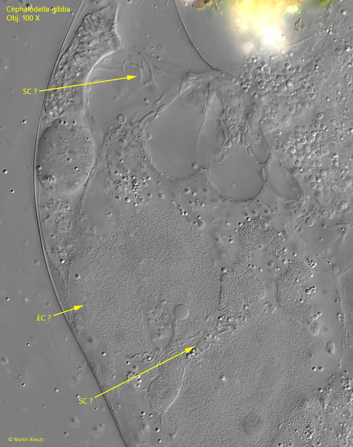
In February 2025, I found a specimen of Cephalodella gibba in the Simmelried with a noticeably enlarged vitellarium, which occupied about two-thirds of the body volume (s. fig. 7). In this vitellarium, conspicuously enlarged cell nuclei were visible. At higher magnification, thread-like structures around these cell nuclei could be seen (s. fig. 8). This might possibly be a phase shortly after fertilization by a male. I have not found any description in the literature about the processes that occur in the vitellarium of monogonont rotifers after sexual fertilization. Therefore, this interpretation is only a hypothesis. However, sperm cells are clearly visible in the strongly compressed specimen, which supports this interpretation (s. fig. 9).
Fig. 7:Cephalodella gibba. A slightly squashed specimen with an enlarged vitellarium and enlarged nuclei (arrows). Obj. 40 X.
Fig. 8:Cephalodella gibba. Possibly this is the vitellarium shortly after fertilization by a male. In that case, the enlarged cell nuclei could be egg cells (EC ?) surrounded by sperm cells (SC ?). Obj. 60 X.
Fig. 9:Cephalodella gibba. In the strongly squasged specimen thread-like structures can be seen very similar to sperm cells (SC ?). EC ? = probably an egg cell. Obj. 100 X.