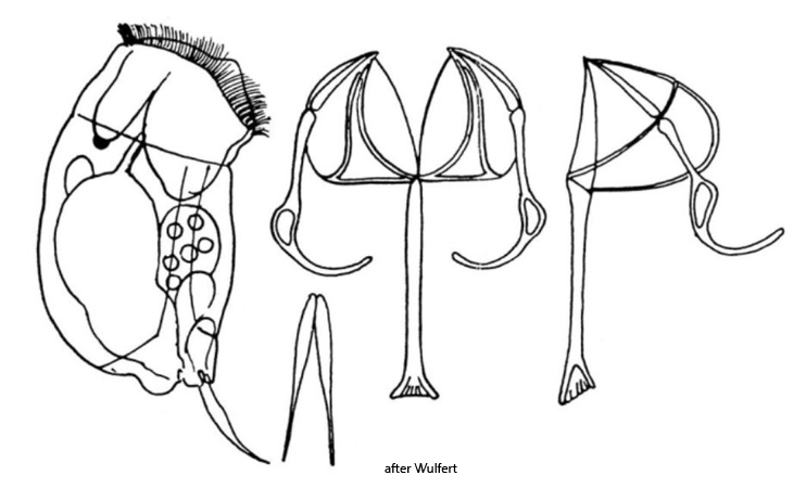
body stout, dorsally curved, ventrally almost straight
length 106-160 µm (with toes)
head slightly separated from body
corona oblique with lips
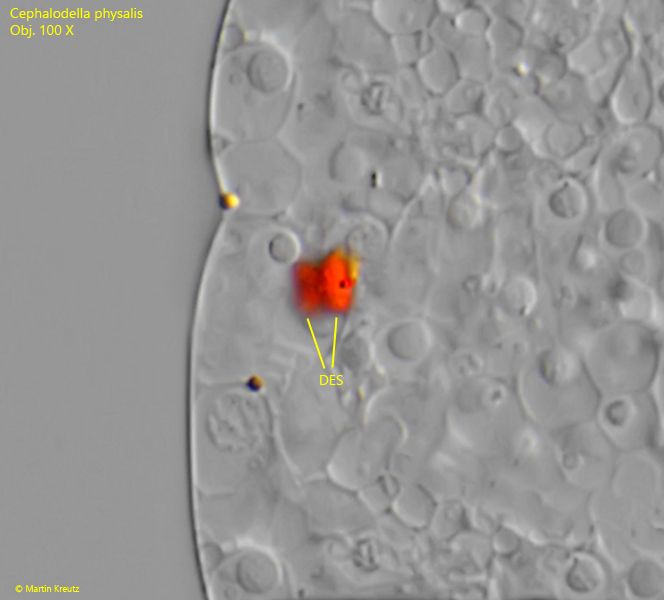
double cervical eyespot
toes slightly thickened in middle, tapered and pointed ends
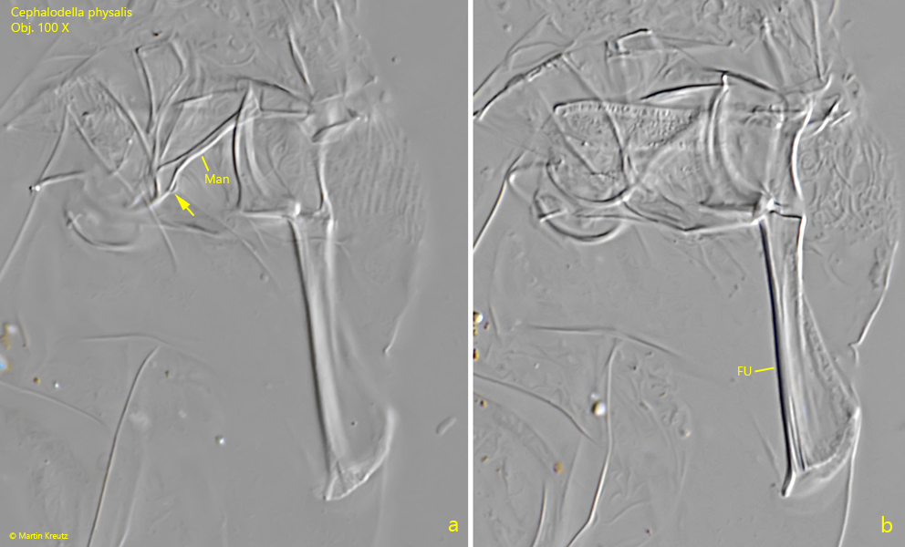
manubria with median loop
Cephalodella physalis
So far, I have only found two specimens of Cephalodella physalis. The first was in November 2019 in the Mühlweiher Litzelstetten and the second in July 2025 in samples from the Schwemm Moor in Austria.
In the samples, Cephalodella physalis stands out due to its stocky body shape, which somewhat resembles Cephalodella auriculata. However, there are some differences that can only be recognized upon closer inspection. The convex dorsal side of Cephalodella physalis appears somewhat humped, not evenly rounded. The toes have a very slight thickening in the middle and then taper off sharply towards the end (s. figs. 2 a and 3 a). They are only slightly bent ventrally. The cervical eyespot is double, which can only be seen in squashed specimens or from dorsal view (s. fig. 4). The exact structure of the trophi is also important, which can only be seen in squashed or macerated specimens. The manubria have a conspicuous loop in the middle (s. fig. 5 a).
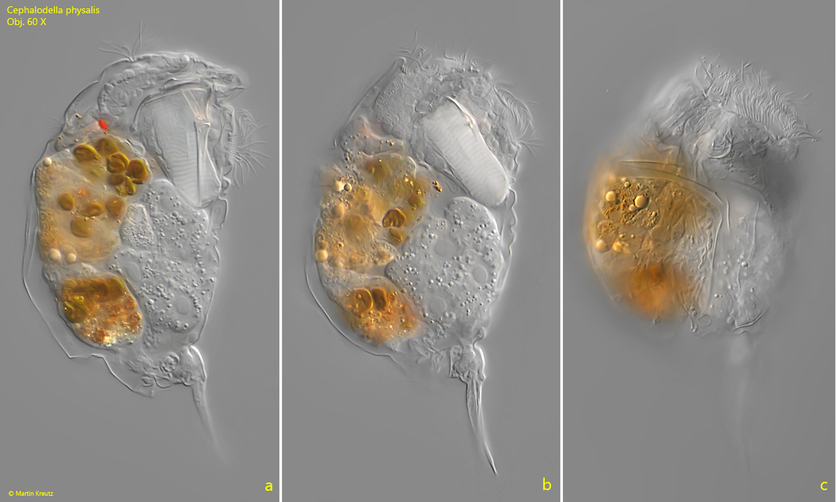
Fig. 1 a-c:Cephalodella physalis. L = 145 µm (with toes). A freely swimming specimen from right. Obj. 60 X.
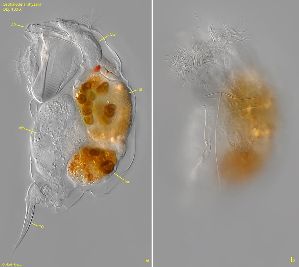
Fig. 2 a-b:Cephalodella physalis. L = 145 µm (with toes). The slightly squashed specimen from left as shown in fig. 1 a-c. Note the extended lips (Lip) of the corona. CG = cerebral ganglion, Int = intenstinum, St = stomach, TO = toes, Vit = vitellarium. Obj. 100 X.
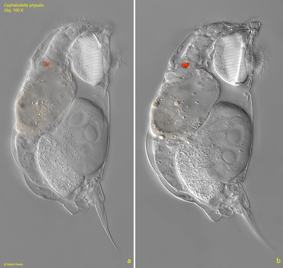
Fig. 3 a-b:Cephalodella physalis. L = 130 µm (with toes). A second specimen from right. Obj. 100 X.
Fig. 4:Cephalodella physalis. The double eyespot (DES) in a strongly sqaushed specimen. Obj. 100 X.
Fig. 5 a-b:Cephalodella physalis. Two focal planes of the trophi. Note the loop (arrow) in the middle of the manubria (Man). Fu = fulcrum. Obj. 100 X.