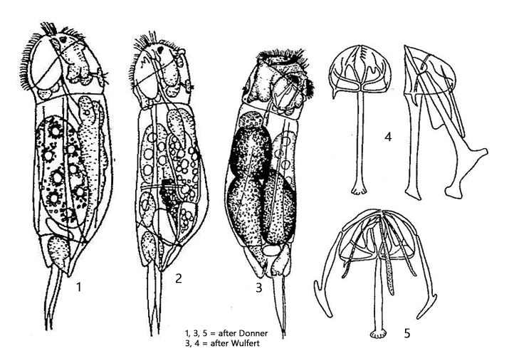
body moderately stout and slightly gibbous dorsally
length 140–250 µm
corona strongly oblique, without lips
ganglion large, saccate
retrocerebral sac sometimes present
neck well marked
lateral clefts nearly parallel-sided.
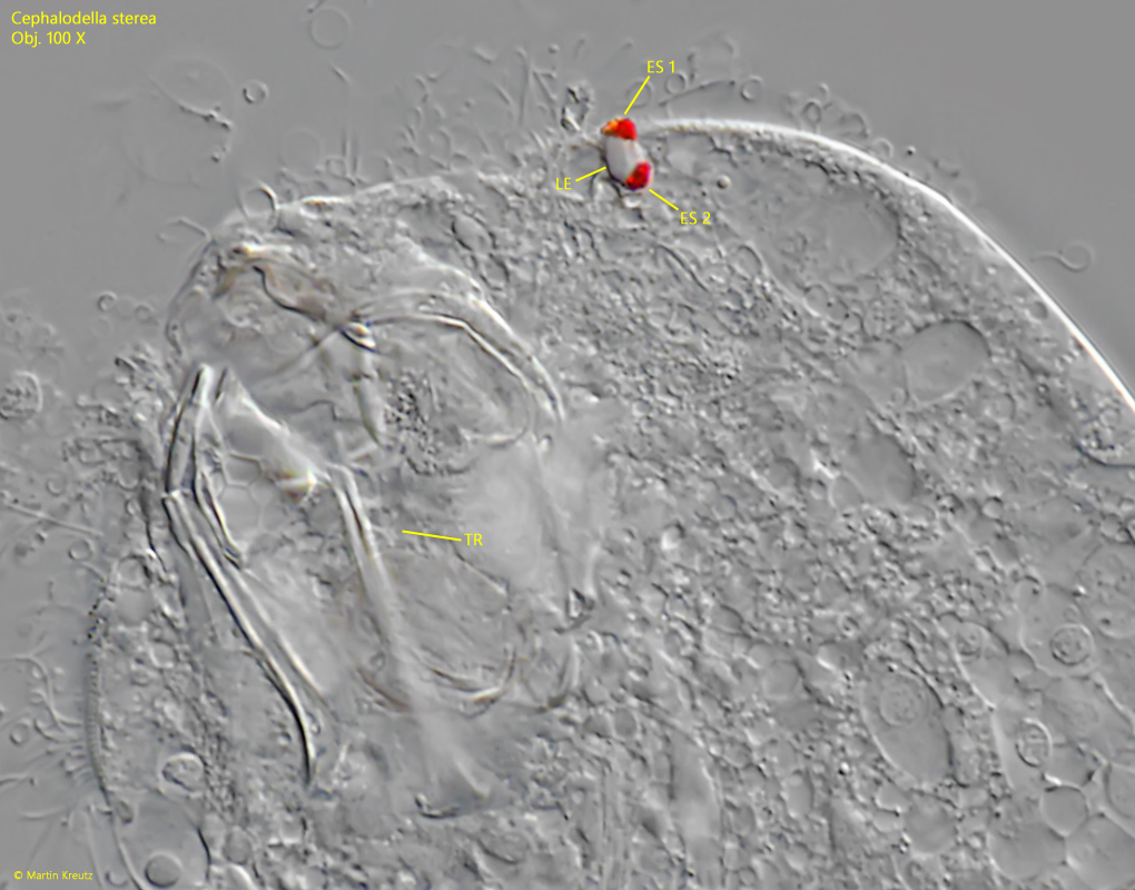
two eyespots attached to one lens
toes are rather short, minutely recurved posteriorly, pointed
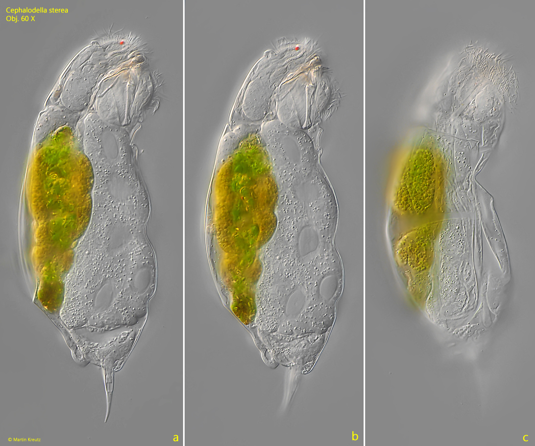
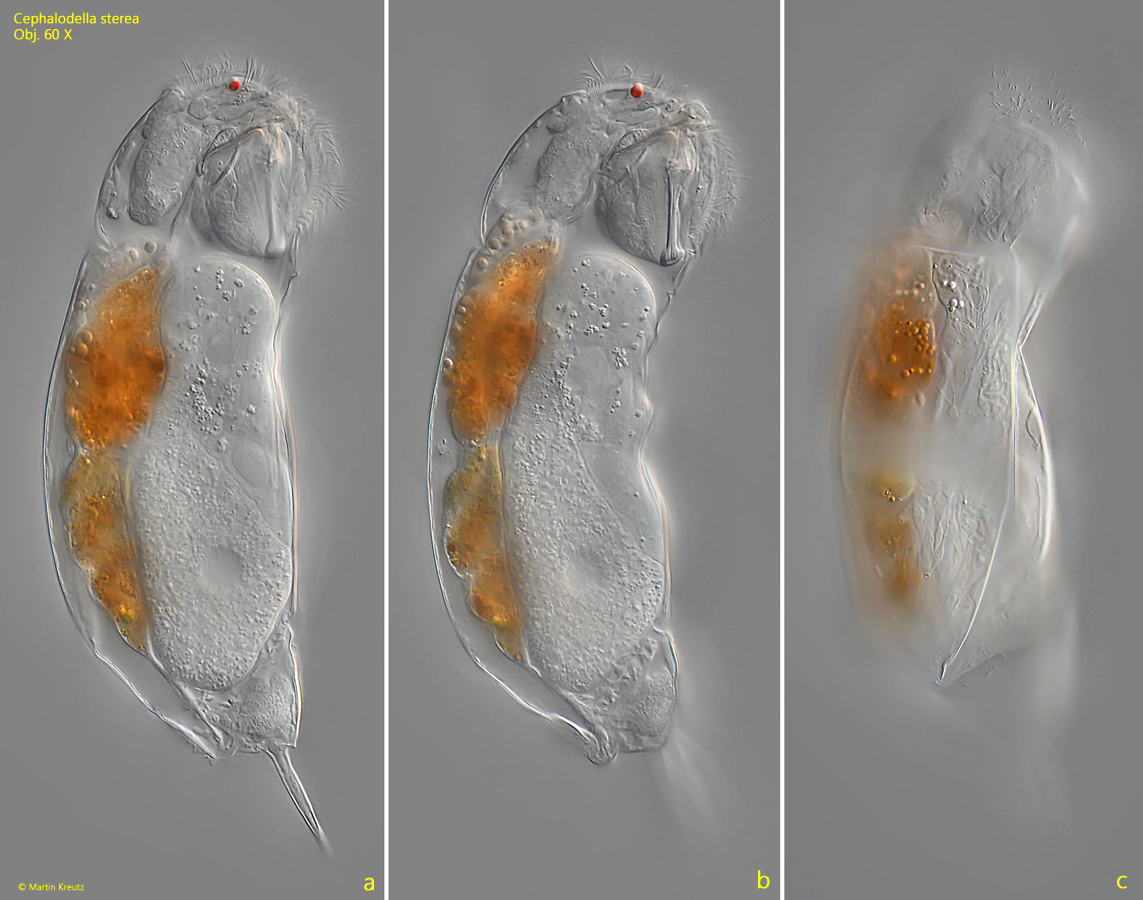
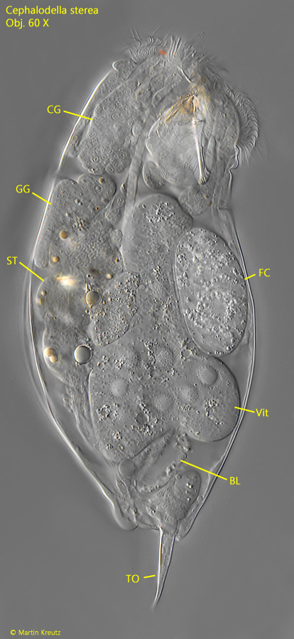
Cephalodella sterea
So far I have only found a few specimens of Cephalodella sterea. In all cases the specimens were found in floating plant masses.
In the samples, the specimens stand out due to their apical eyespot. This is particularly structured, as two closely spaced eyespots are attached to a common lens. However, this special arrangement can only be recognized in heavily squashed specimens (s. fig. 4).
Another important characteristic of Cephalodella sterea is the shape of the toes. They taper to a sharp point and are slightly bent backwards at the end (s. fig. 1 a). The length of the toes is variable in this species.
I have also found a few specimens which were infested by the parasitic fungus Bertramia aspeospora (s. fig. 3). This fungus attacks many species of rotifers and is more common. The very large fungal cell is located in the body cavity. After multiple cell divisions, flagellated spores are formed which are released from the now dead host. The life cycle of Bertramia aspeosporawas studied in detail by Plewka.
Fig. 1 a-c:Cephalodella sterea. L = 172 µm. A freely swimming specimen from right. Obj. 60 X.
Fig. 2 a-c:Cephalodella sterea. L = 172 µm. A second specimen from right. Obj. 60 X.
Fig. 3:Cephalodella sterea. L = 162 µm. A slightly squashed specimen infested by the parasitic fungus Bertramia aspeospora. The fungal cell (FC) is large and filled with refractive spherules. BL = bladder, CG = cerebral ganglion, GG = gastric gland, ST = stomach, TO = toes, Vit = vitellarium. Obj. 60 X.
Fig. 4:Cephalodella sterea. Apically two eyespots (ES 1, ES 2) are attached to one lens (LE). TR = trophi. Obj. 100 X.
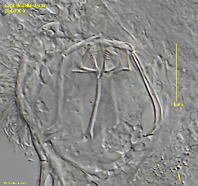
Fig. 5:Cephalodella sterea. The trophi in a strongly squashed specimen. Obj. 100 X.