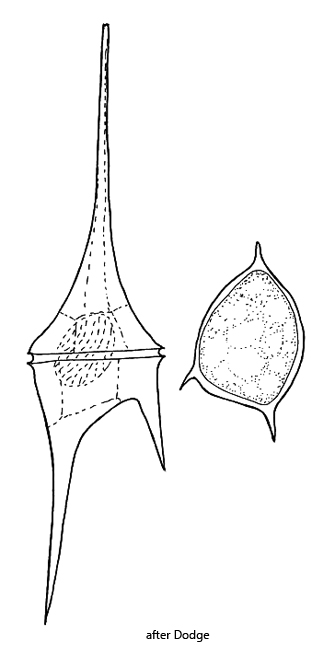
Ceratium furcoides
(Levander) Langhans, 1925
Most likely ID: n.a.
Â
Synonym: Ceratium hirundinella var. furcoides, Ceratium hirundinella f. furcoides, Ceratium furca var. lacustre, Ceratium furcoides f. gracile
Â
Sampling location: Lake Constance, Mühlhalden pond, Hagstaffel pond, Mühlweiher Litzelstetten, Pond of the waste disposal company Constance.
Â
Phylogenetic tree: Ceratium furcoides
Â
Diagnosis:Â
- cells elongated pyramidal or spindle-shaped
- length 123–300 µm, width 28–42 µm
- epitheca a long, slender horn
- hypotheca with 2 horns of different length
- sometimes hypotheca with third rudimentary horn
- theca arranged of several plates with reticulate ornamentation
- numerous yellowish-brown or greenish-orange chloroplasts
- oval nucleus located in epitheca
- two flagella (transversal and longitudinal)
Â
The epitheca and the hypotheca are separated by a transverse groove called the cingulum (s. fig. 2 a-b). The transverse flagellum runs in it, performing a wavelike movement there (s.fig. 4). In freely swimming specimens, it is difficult to see. A second, much longer flagellum originates from the longitudinal sulcus, which has a complex structure in Ceratium furcoides. This longitudinal flagellum extends beyond the posterior horn and provides propulsion (s. fig. 3 a-b).
Â
The theca of Ceratium furcoides consists of cellulose and is composed of 16 plates. The plates exhibit a net-like pattern of depressions. In the area of the sulcus, there is a region where the two flagella originate.
Ceratium furcoides can eject very delicate, needle-shaped extrusomes. These originate from the contents of the Golgi apparatuses, which are distributed in the cytoplasm and can release their contents (Leadbeater & Düdge, 1967). I was also able to observe these extrusomes, as they are ejected en masse in heavily compressed specimens (s. fig. 6). They are about 10–40 µm long.
Fig. 1 a-d: Ceratium furcoides. L = 297 µm. Different focal planes of a freely swimming specimen from ventral. Obj. 40 X.
Fig. 2 a-b: Ceratium furcoides. L = 297 µm. The same specimen as shown in fig. 1 a-d at higher magnification. The anterior epitheca (EP) is separated by the transverse cingulum (CI) separated from the posterior hypotheca (HY). In the epitheca the nucleus (Nu) is located. The sulcus (SU) is a longitudinal, ventral furrow. Obj. 40 X.
Fig. 3 a-b: Ceratium furcoides. L = 312 µm. A second specimen from dorsal with focal plane on the cingulum (CI) and on the nucleus (Nu). Posterior the longitudinal flagellum (LF) is visible. Obj. 60 X.
Fig. 4: Ceratium furcoides. In the furrow of the cingulum the transverse flagellum (TF) is visible in a wavy movement. Chl = chloroplasts, LF = longitudinal flagellum. Obj. 100 X.
Fig. 5: Ceratium furcoides. Focal plane on the longitudinal furrow of the sulcus (SU) from dorsal. The longitudinal flagellum (LF) arise from the sulcus. Nu = nucleus. Obj. 60 X.
Fig. 6: Ceratium furcoides. In a strongly squashed specimen the fine, needle-shaped extrusomes (EX) become visible. They have a length of about 10–40 µm. Obj. 100 X.