I find Ceratium hirundinella exclusively in the plankton of Lake Constance. In contrast, at my other sampling sites—which are smaller bodies of water—only the similar species Ceratium furcoides was present.
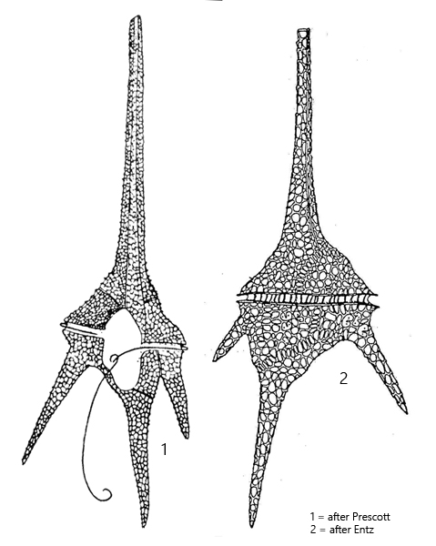
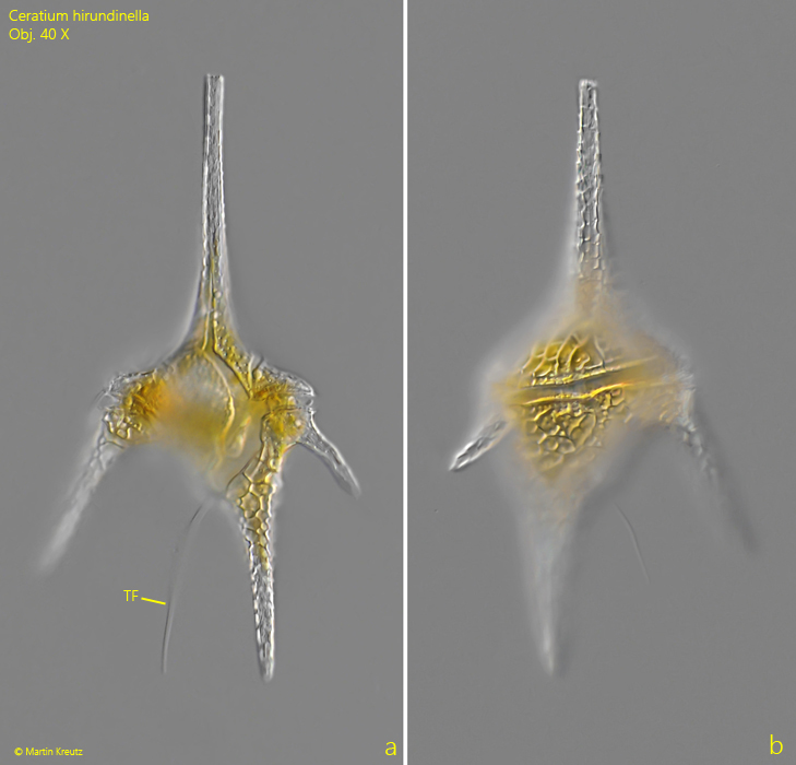
The specimens of Ceratium hirundinella in my population were mostly between 170 and 210 µm in length. Unlike Ceratium furcoides, Ceratium hirundinella exhibits a pronounced shoulder formation at the transition from the epitheca to the hypotheca (s. fig. 1 a-b). In addition, the antapical horns of the hypotheca are more widely spread than in Ceratium furcoides.
Another difference, though more difficult to discern, concerns the structure of the epitheca. In both species, the epitheca is composed of four plates. In Ceratium hirundinella, all four plates extend to the tip of the apical horn. In Ceratium furcoides, however, only three plates extend to the tip. However, this characteristic is very difficult to discern in living specimens. Therefore, the number of horns and the body shape are decisive. In Ceratium hirundinella, the hypotheca has three horns that form a wide angle with one another, whereas Ceratium furcoides has only two horns, with the longer one extending almost straight backward. Only rarely does a rudimentary, very small third horn develop in Ceratium furcoides. As a result, Ceratium furcoides has a slender, elongated body shape, while Ceratium hirundinella appears much more expansive and stocky.
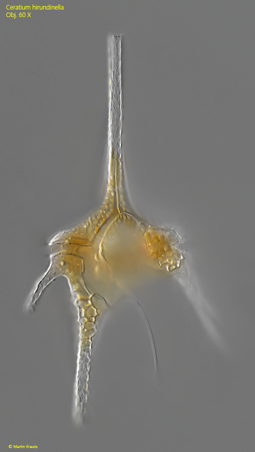
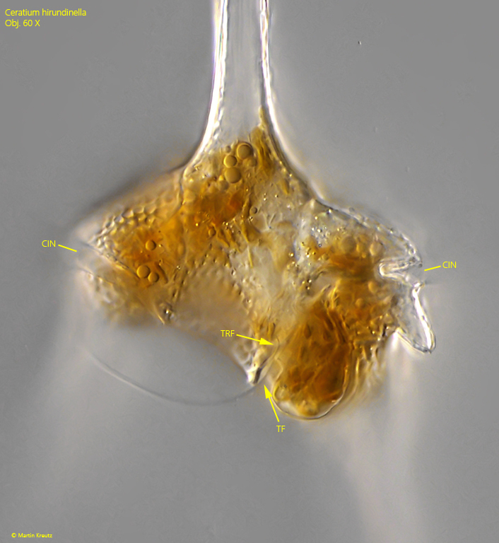
The epitheca and the hypotheca are separated by a transverse groove called the cingulum. The transverse flagellum runs through this groove, where it performs a wave-like motion. In free-swimming specimens, it is difficult to see. A second, much longer trailing flagellum originates from the longitudinal sulcus, which has a complex structure in Ceratium hirundinella. This longitudinal trailing flagellum extends beyond the posterior horn and provides propulsion.
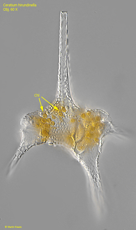
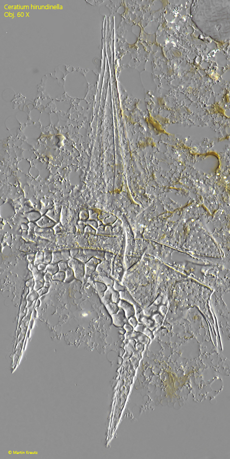
The theca of Ceratium hirundinella consists of cellulose and is composed of 16 plates. The plates exhibit a net-like pattern of depressions. In the region of the sulcus, there is an area where the two flagella emerge from a gap between the plates (s. fig. 5).
Fig. 1 a-b:Ceratium hirundinella. L = 194 µm. A freely swimming specimen from ventral (a) and dorsal (b). TF = trailing flagellum. Obj. 40 X.
Fig. 2:Ceratium hirundinella. L = 194 µm. The same specimen as shown in fig. 1 a-b at higher magnification from ventral. Obj. 60 X.
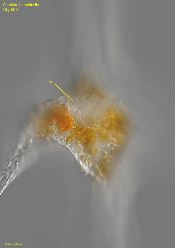
Fig. 3:Ceratium hirundinella. The large nucleus (Nu) is located beneath the apical horn. Obj. 60 X.
Fig. 4:Ceratium hirundinella. The disc-shaped chloroplasts (Chl) in a slightly squashed specimen. Obj. 60 X.
Fig. 5:Ceratium hirundinella. The transverse flagellum (TRF) in the cingulum (CIN) as well as the trailing flagellum (TF) arise from a ventral slit between two plates. Obj. 60 X.
Fig. 6:Ceratium hirundinella. The reticulate pattern of the theca covering the body in a strongly squashed specimen. Obj. 60 X.