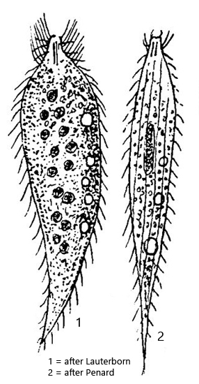
So far, I have only been able to find one specimen of Chaenea limicola in December 2025 in the mud of the Simmelried. The species seems to be generally rare, as there are very few reports on Chaenea limicola. Apart from Lauterborn (1901) and Penard (1922), no other observations seem to exist. Kahl (1935) only provides a brief description of these two authors.
My specimen was somewhat larger at 192 µm in length than those found by Lauterborn and Penard. I was able to observe the specimen freely swimming in its natural form (s. fig. 1 a-d). As described by Penard, the cone-shaped oral bulge is almost always held at an angle while swimming. The entire body is slender spindle-shaped and tapers to a posterior point.
The contractile vacuoles are very clearly visible, arranged in a longitudinal row. I could identify at least 4 contractile vacuoles, which decrease in size towards the posterior end (s. fig. 1 b). I could not observe that the rearmost contractile vacuole is the largest, as described by Penard.
The cytoplasm is filled with ring-shaped granules, which makes it appear yellowish and opaque. This ring-shaped granule was also observed by Lauterborn. It possibly originates from the prey (e.g., Loxocephalus). The numerous nodes of the macronucleus, which are scattered in the cytoplasm, can only be recognized in the strongly squashed specimen (s. fig. 3). Although Penard’s drawing suggests that there is only a single central, ellipsoid macronucleus, he describes it as a concentrated collection of macronuclear nodules. His observations of the nuclear apparatus are therefore consistent with those of Lauterborn.
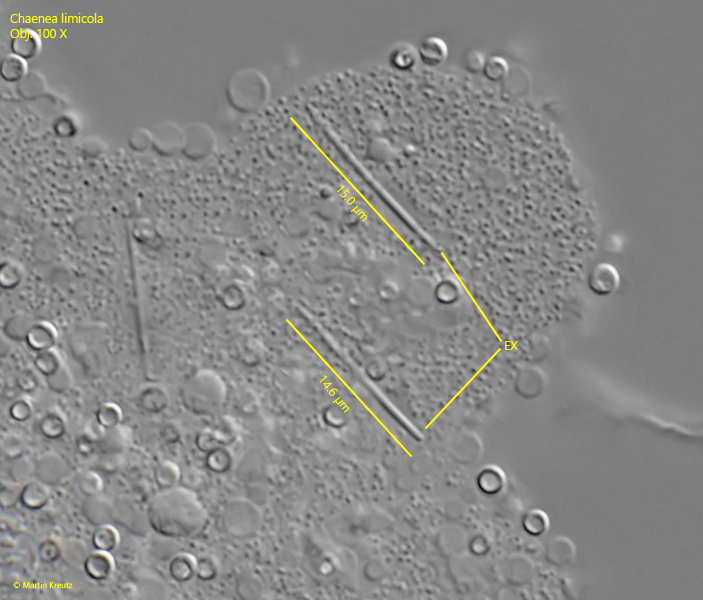
The rod-shaped extrusomes of Chaenea limicola are not described in detail by earlier authors. According to my measurements, they are 14–15 µm long (s. fig. 4). In the pharynx, only a few of these extrusomes are arranged in a bundle. The remaining extrusomes are scattered throughout the cytoplasm.
Fig. 1 a-d: Chaenea limicola. L = 192 µm. A freely swimming specimen. Note the several contractile vacuoles (arrows) arranged in a longitudinal row. The cone-shaped oral bulge is always hold crooked. Obj. 40 X.
Fig. 2 a-e: Chaenea limicola. L = 192 µm. The same specimen as shown in fig. 1 a-d at higher magnification. The dorsal brush (DB) ist visible. The cytoplasm is completely filled with ring-shaped granules (likely from prey). Obj. 60 X.
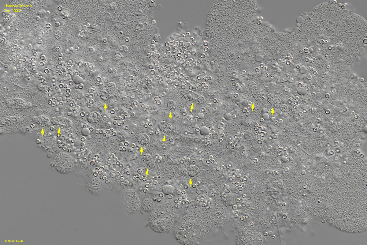
Fig. 3: Chaenea limicola. In the strongly squashed specimen the numerous nodules of the macronucleus are visible (arrows). Obj. 100 X.
Fig. 4: Chaenea limicola. The rod-shaped extrusomes (EX) have a length of 14–15 µm. Obj. 40 X.