gelatinous sheath limited by distinct membrane in young cells, otherwise amorphous
cells ovoid or oval, length 5–14 µm
one chloroplast, multiple lobed
one pyrenoid, localized in one of the lobes
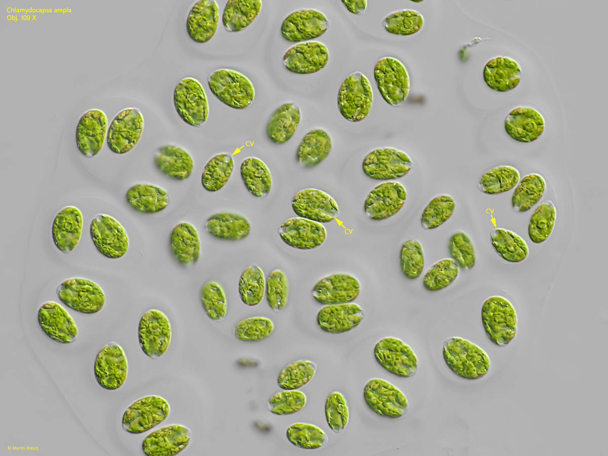
contractile vacuole in the narrowed cell end
one eyespot (hard to see)
vegetative cells can form flagella
Chlamydocapsa ampla
I find Chlamydocapsa ampla regularly but rarely in two of my sampling sites. Mostly I find the colonies growing on the surface of the mud layer.
The main characteristics of Chlamydocapsa ampla are the mostly ovoid shaped cells, which are located in a gelatinous sphere with a distinct, sharply defined membrane. There are 2–8 cells in each of these gelatinous spheres. These spherical colonies are often connected to form larger, irregularly shaped aggregates, which then appear grape-shaped (s. fig. 1 a-b).
Chlamydocapsa ampla belongs to the tetrasporal algae, which have a contractile vacuole and an eyespot. In addition, the vegetative cells can form a pair of flagella, although this is rarely observed. The contractile vacuole is located in the tapered end of the cell, which is often transparent (s. figs 2 and 4). The eyespot is adhering to the cell wall, is very small and can only be seen at high magnification (s. figs. 2 and 4).
Fig. 1 a-b: Chlamydocapsa ampla. L = 8–12 µm (cells). Two focal planes of a colony with a diameter of 200 µm. Obj. 40 X.
Fig. 2: Chlamydocapsa ampla. L = 8–12 µm (cells). A detail of the colony as shown in fig. 1 a-b. Note the contractile vacuoles (CV) and eyespots (ES) in the cells. Nu = nucleus, PY = pyrenoid. Obj. 100 X.
Fig. 3: Chlamydocapsa ampla. D = 20–24 µm (of colonies). An aggregation of young, spherical colonies with each 2 or 4 cells. Obj. 40 X.
Fig. 4: Chlamydocapsa ampla. L = 7–13 µm (of cells). Some colonies as shown in fig. 3 in detail. The cells are ovoid. The contracile vacuole (CV) in located in the tapered end of the cells. ES = eyespot, SM = spherical membrane. Obj. 100 X.
Fig. 5: Chlamydocapsa ampla. D = 62 µm (of colony). A young, spherical colony. Obj. 100 X.
Fig. 6: Chlamydocapsa ampla. L = 12–15 µm (of cells). Cells in a squashed colony. CV = apical contractile vacuoles. Obj. 100 X.