The habitat of Chlamydomyxa labyrinthuloides is bogs and the detritus of ponds. I myself have so far only found Chlamydomyxa labyrinthuloides in the Schwemm in Austria and once in the pond of the waste disposal company Constance. These locations are completely different, as the Schwemm is a largely untouched raised bog, while the pond of the waste disposal company Constance is a heavily eutrophic, artificially created body of water. The specimen shown below comes from the Schwemm, where I found a few specimens in June 2026.
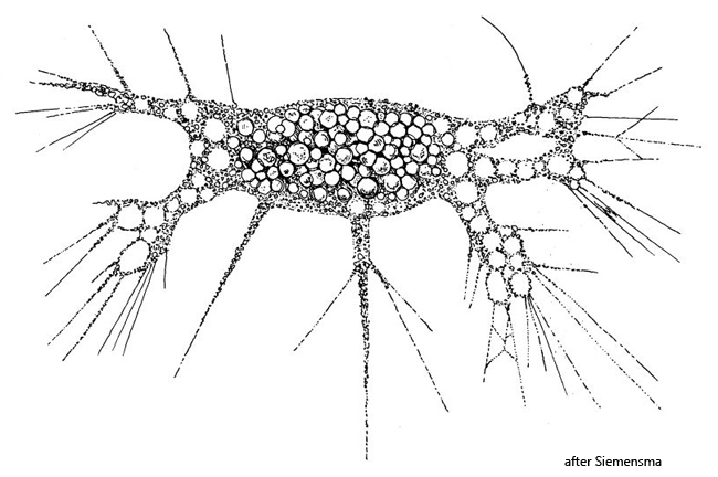
In 1904, Cash described an amoeba which he called Penardia mutabilis. In 1991, the species was thoroughly examined by Page and Siemensma, and they found very strong similarities with Chlamydomyxa labyrinthuloides, an amoeba described by Archer in 1875. Therefore, Penardia mutabilis is now considered a junior synonym of Chlamydomyxa labyrinthuloides. The name given to a species first has priority.
Based on morphological and genetic studies, Chlamydomyxa labyrinthuloides is now classified among the heterokonts and is closely related to the chrysophytes. This classification is based, among other things, on the composition of the chlorophylls and pigments, which are responsible for the brownish-yellow coloration of the chloroplasts.
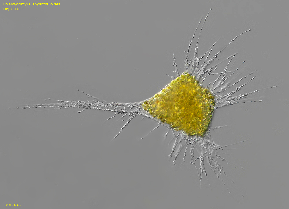
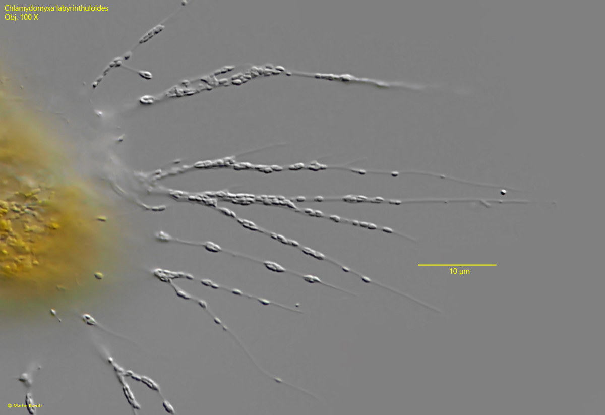
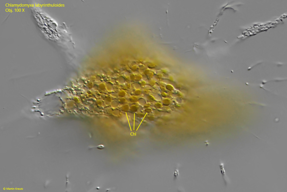
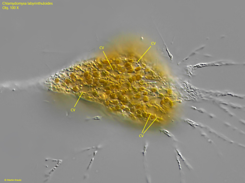
Characteristic of Chlamydomyxa labyrinthuloides is the yellowish-green color of the cytoplasm and the strongly granulated filopodia. At high magnification, it can be seen that these granules are ellipsoid in shape, with a length of 0.8–1.2 µm (s. fig. 2). Because of this shape, they resemble bacteria. The chloroplasts can be clearly seen in the cytoplasm as yellow-brown discs with a diameter of 1.9–2.3, as they are also located in the periphery (s. fig. 3). The numerous contractile vacuoles near the cell surface are also visible (s. fig. 4). I was unable to examine the cell nuclei, as the layer thickness could not be further reduced due to foreign bodies under the coverslip.
Fig. 1: Chlamydomyxalabyrinthuloides. L = 250 µm. A fully extended and moving specimen. Obj. 60 X.
Fig. 2: Chlamydomyxalabyrinthuloides. The filopodia in detail with the elliptically shaped granules. The granules have a length of 0.8–1.2 µm. Obj. 100 X.
Fig. 3: Chlamydomyxalabyrinthuloides. Focal plane on the disc-shaped chloroplasts (Chl) scattered in the cytoplasm. They have a diameter of 1.9–2.3 µm. Obj. 100 X.
Fig. 4: Chlamydomyxalabyrinthuloides. The small contractile vacuoles (CV) near the cell surface. Obj. 100 X.