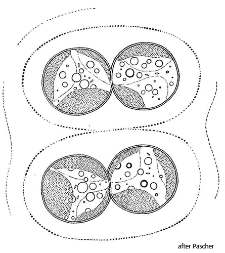
fragments of mother cell wall in mucilaginous envelope
1–4 parietal chloroplasts, pale yellowish green
chloroplasts disc-shaped or cup-shaped, in older cells diffuse
prominent red or orange oil droplet present
Chlorobotrys regularis
I regularly find Chlorobotrys regularis in the Simmelried. This Xanthophycee is not present in my other sampling sites.
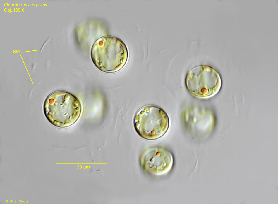
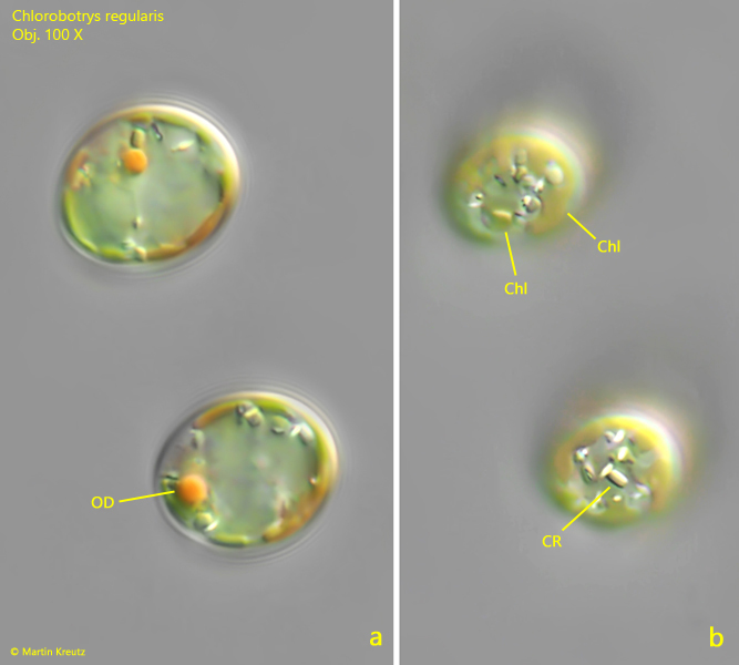
I recognize Chlorobotrys regularis mainly by the yellow-green chloroplasts and the typical orange or red oil drop that can be found in most of the cells (s. fig. 4). The older cells are always spherical with a thick cell wall. They lie in a mucilaginous sheath, which also contains the remains of the mother cell wall (s. fig. 3). In my population, the cells were never larger than 16 µm.
The similar species Chlorobotrys polychloris has considerably more chloroplasts and the cells are also significantly larger at 18–25 µm.
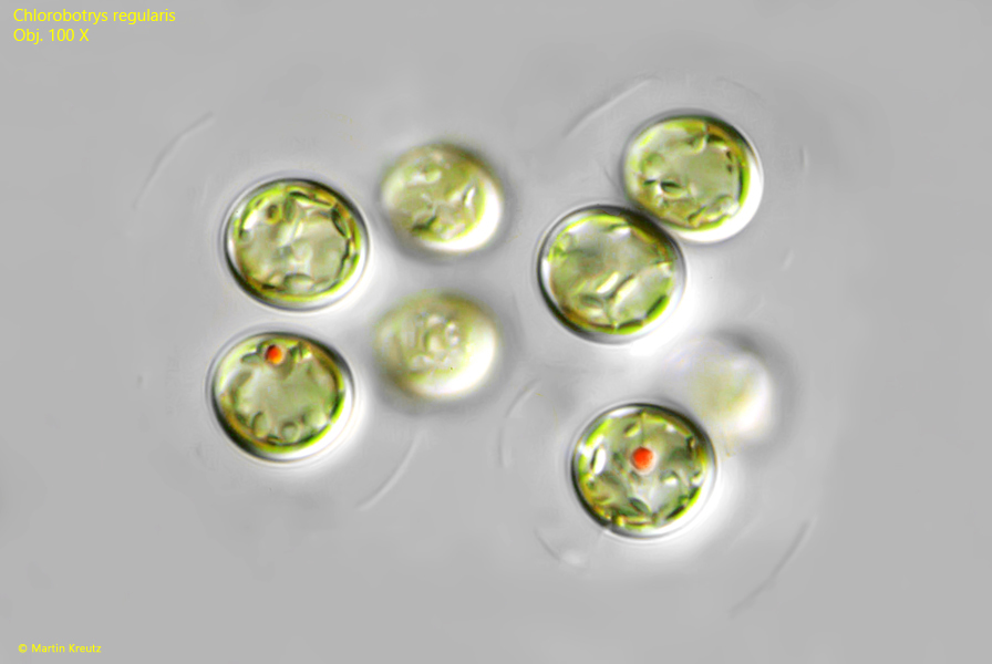
Fig. 1:Chlorobotrys regularis. D = 14–16 µm (of cells). A group of 8 cells. Obj. 100 X.
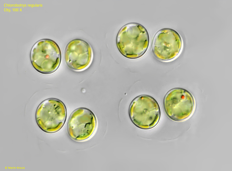
Fig. 2:Chlorobotrys regularis. D = 13–15 µm (of cells). A second group of 8 cells. Obj. 100 X.
Fig. 3:Chlorobotrys regularis. D = 14–15 µm (of cells). A third group of 8 cells. RM = remains of mother cell. Obj. 100 X.
Fig. 4:Chlorobotrys regularis. D = 14–15 µm (of cells). Two focal planes of two young cells. Note the distinct, orange colored oil droplet (OD). Chl = parietal chloroplasts, CR = crystals. Obj. 100 X.