Chlorochromatium aggregatum
Lauterborn, 1906
Most likely ID: n.a.
Â
Synonym: n.a.
Â
Sampling location: Main bike path pond
Â
Phylogenetic tree: n.a.
Â
Diagnosis:Â
- consortium of phototroph sulfur bacterium and heterotroph bacterium
- consortia ellipsoid, oval or cyndrical, green-yellowish colored
- consortica often with equatorial constriction
- one central heterotroph bacterium covered by 10–16 green sulfur bacteria
- length (of consortia) 9–12 µm, width 5–7 µm
- sulfur bacteria green-yellowish, flagellum absent
- heterotroph bacterium colorless, one flagellum
I have only found Chlorochromatium aggregatum once so far, in March 2026 in a small pond on the Main bike path near Würzburg. The pond is heavily eutrophicated with a black, anaerobic sludge layer in shallow water. In the samples from this sludge layer, Chlorochromatium aggregatum was found in large quantities.
Â
Chlorochromatium aggregatum was first described in 1906 by Lauterborn. In 1914, Bude recognized that it is a consortium of green sulfur bacteria attached to a central, colorless bacterium with a flagellum.
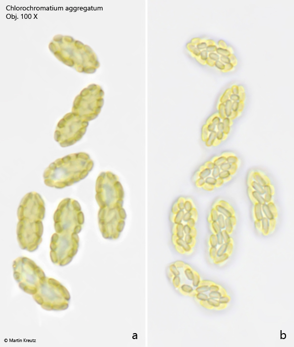
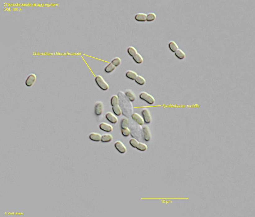
The epibiotic green sulfur bacterium was identified as Chlorobium chlorochromatium. This bacterium is capable of photosynthesis. The green-yellow coloration is caused by bacteriochlorophylls a and c (Imhoff, 1995). In culture experiments, it was demonstrated that Chlorobium chlorochromatium can be isolated and can also grow in culture without the central bacterium. Chlorobium chlorochromatium is strictly anaerobic. It uses sulfides as electron donors to fix CO2 and N2.
Â
The central bacterium was identified by Liu et al. (2013) as Symbiobacter mobilis (referred to by the authors as “Candidatus Symbiobacter mobilis”). It possesses a polar flagellum, which makes the entire consortium motile. Symbiobacter mobilis cannot be isolated and cultured. The genome of this bacterium is so reduced that it can only survive with the help of its epibionts. It uses the photosynthetically produced products of the epibionts for energy generation. For this, Symbiobacter mobilis requires an electron acceptor. Liu et al. (2013) postulate that quinones are delivered to Symbiobacter mobilis by the epibionts, which are reduced there to hydroquinones. These are then returned to the epibionts. Thus, it is an electron cycle system.
Fig. 1: Chlorochromatium aggregatum. Overview of an accumulation of specimens around a filament of Oscillatoria spec. Obj. 60 X.
Fig. 2 a-b: Chlorochromatium aggregatum. Two focal planes of an accumulation of specimens around a detritus and mineral grains. Obj. 100 X.
Fig. 3 a-b: Chlorochromatium aggregatum. Two focal planes of some specimens in DIC. The green-yellowish colored sulfur bacteria Chlorobium chlorochromatii cover the central, colorless and heterotroph bacterium Symbiobacter mobilis. Obj. 100 X.
Fig. 4 a-b: Chlorochromatium aggregatum. The same specimens as shown in fig. 3 a-b in brightfield illumination. Obj. 100 X.
Fig. 5: Chlorochromatium aggregatum. After few minutes under the coverslip the green sulfur bacteria Chlorobium chlorochromatii begin to detach from the central bacterium and the shape of Symbiobacter mobilis becomes visible. Obj. 100 X.