apices broadly rounded, sometimes thickened internally
girdle bands absent
terminal vacuoles with 10–30 small gypsum crystals
Closterium lunula
So far I have only found Closterium lunula in the Paradieswiesen in Austria. I have not yet been able to find this species in the sites in my immediate vicinity.
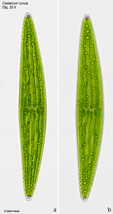
The cells of Closterium lunula are very large and stand out due to their almost straight shape, which differs from the crescent shape of other Closterium species. Numerous pyrenoids are scattered throughout the two chloroplasts (s. fig. 4). The ridges of the chloroplasts are clearly notched, which is very typical for this species (s. fig. 3 b). The terminal vacuoles in my population were filled with a fine crystal grit, with only a few larger crystals (s. fig. 5). The very fine and inconspicuous striation of the cell wall can only be recognized at highest magnification (s. fig. 6).
Fig. 1 a-b:Closterium lunula. L = 575 µm. Two focal planes of a slightly squashed specimen. Obj. 20 X.
Fig. 2 a-b:Closterium lunula. L = 575 µm. The same specimen as shown in fig. 1 a-b in brightfield illumination. Obj. 20 X.
Fig. 3 a-b: A semi-cell in detail. Note the notched ridges of the chloroplast (arrows). Obj. 40 X.
Fig. 4:Closterium lunula. The scattered pyrenoids (PY) in the chloroplasts. The pyrenoids are covered with a layer of starch grains (SG). Obj. 100 X.
Fig. 5:Closterium lunula. The terminal vacuole (TV) in the apex of this specimen is filled with crysal grit. Obj. 100 X.
Fig. 6:Closterium lunula. The striation of the cell wall is very fine and inconspicuous. This specimen has 19 stria/10 µm. Obj. 100 X.