terminal vacuoles with single or few small crystals
nucleus central
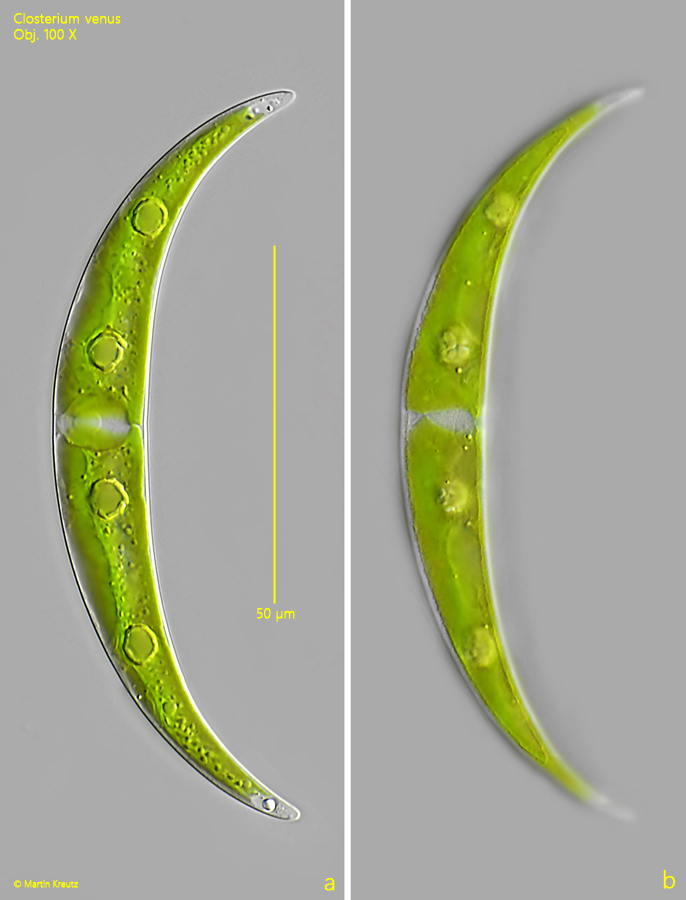
Closterium venus
I find Closterium venus quite frequently, but always only single specimens. The most important distinguishing feature is the small size of 100 µm or less, as well as the slender and uniformly curved shape.
In my population the cells had always 2 pyrenids per semi-cell. This corresponds to the description of Closterium venus by Förster (1982). I have never found cells with more pyrenoids. Lenzenweger (1996), however, describes the species with “some” pyrenoids and draws it with 6 pyrenoids per semi-cell (s. drawing above).
The differentiation from the very similar species Closterium incurvum is difficult. The cells of this species have an almost semicircular curvature and the apices are very pointed. In addition, the cells of Closterium incurvum are a maximum of 80 µm long and therefore somewhat smaller than those of Closterium venus. Otherwise, the characteristics are identical. Since the cells of my population are significantly less curved and have a length of about 100 µm, they can be assigned to Closterium venus.