dorsal part of shell covered with spines (10–14 µm)
one nucleus, sometimes with central nucleolus
cytoplasm sometimes with symbiotic algae
3 or more contractile vacuoles
slender pseudopodia, about twice body length
Cochliopodium vestitum
I find Cochliopodium vestitum very frequently in my samples. Very often, they crawl on detritus flakes or gelatinous bacterial flakes. This amoeba can also be detected for a long time in old samples. It often settles on the floating coverslip to graze on algae and bacteria.
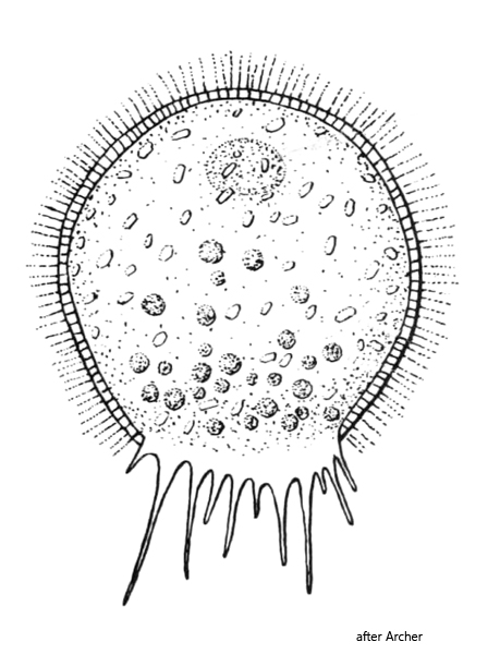
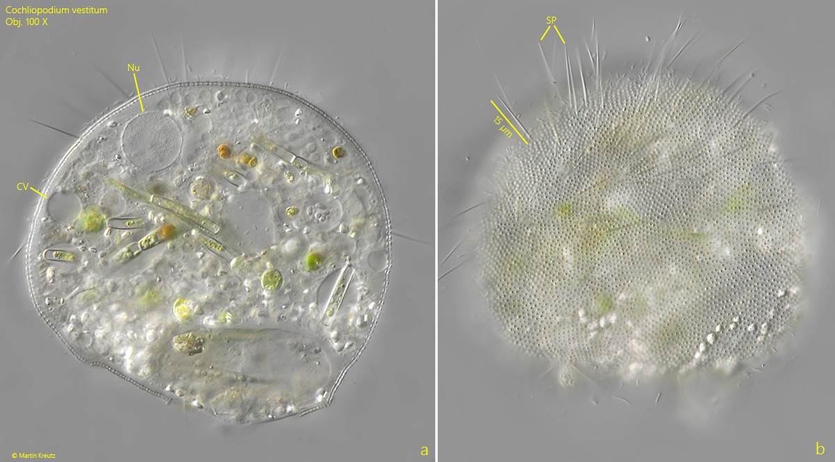
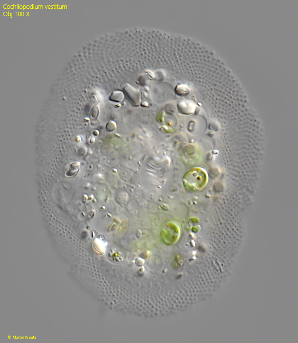
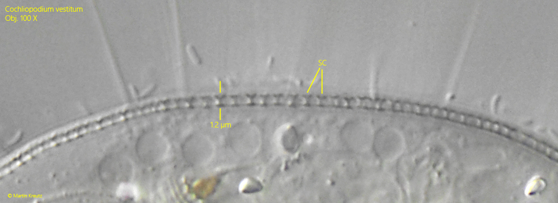
Cochliopodium vestitum was first described by Archer (1871). Although the species is very common, it was not redescribed in detail until 2005 by Kudryavtsev. The body of Cochliopodium vestitum is usually approximately round in shape and enclosed by a membranous, flexible shell. This shell is covered with very small scales that appear as granules when viewed from above and are T-shaped in lateral view at the highest magnification (s. fig. 7). In fact, these scales are very complex in structure, which can only be seen under a scanning electron microscope. In addition to these scales, spines approximately 15 µm long are attached to the shell. These are located primarily on the posterior half of the shell (s. figs. 3 b).
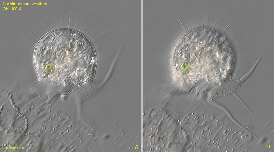
The mouth opening is flexible and has no fixed shape. However, it can be identified by the scales on the shell. The extended pseudopodia are slender and taper at the end (s. fig. 4 a-b). According to my observations, they reach a maximum length of twice the body length.
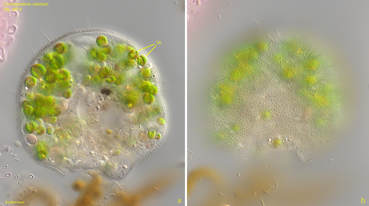
The cytoplasm is usually filled with phagocytosed algae, purple bacteria, or even euglenoids (s. fig. 3 a). Starch grains are often found in the cytoplasm. In addition to the colorless form, specimens containing symbiotic algae are also often found (s. fig. 6 a-b). These have a cup-shaped chloroplast and are of the Chlorella type.
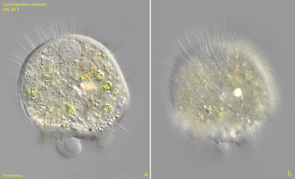
Fig. 1 a-b:Cochliopodium vestitum. D = 75 µm. Two focal planes of a slightly squashed specimen with a yellowish coloration. Obj. 60 X.
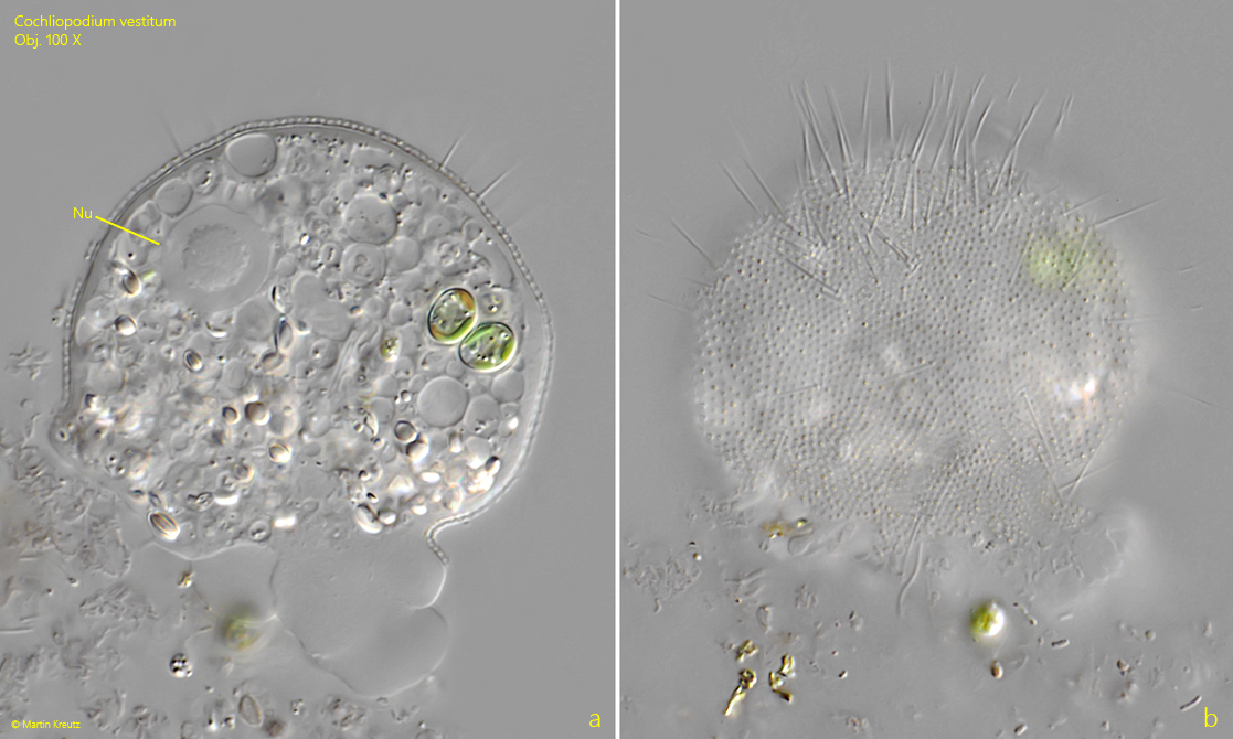
Fig. 2 a-b:Cochliopodium vestitum. D = 57 µm. A second slightly squashed specimen. Note the nucleus with a central nucleolus. Obj. 100 X.
Fig. 3 a-b:Cochliopodium vestitum. Two focal planes of a squashed specimen. The spines (SP) with a length of about 15 µm are mainly located on the dorsal part of the shell. CV = one of the contractile vacuoles, Nu = nucleus. Obj. 100 X.
Fig. 4 a-b:Cochliopodium vestitum. D = 44 µm. A specimen with elongated pseudopodia. The pseudopodia are slender and tapered to the distal end. Obj. 100 X.
Fig. 5:Cochliopodium vestitum. D = 70 µm. A specimen in ventral view gliding along the coverslip. The scales appear as granules. Obj. 100 X.
Fig. 6 a-b:Cochliopodium vestitum. D = 62 µm. A specimen with symbiotic algae (SA). The algae appear to be of the Chlorella type. Obj. 100 X.
Fig. 7:Cochliopodium vestitum. The scales (SC) are 1.2 µm high and have a T-shape in lateral view. The complex fine structure of the scales cannot be resolved in the light microscope. Obj. 100 X.