Codonella cratera

(Leidy, 1877) Imhof, 1885

Most likely ID: n.a.


Synonym: Tintinnopsis lacustris


Sampling location: Mühlhalden pond, Lake Constance, Hagstaffel pond, Pond of the waste disposal company Constance, Mühlweiher Litzelstetten


Phylogenetic tree: Codonella cratera


Diagnosis: 

  • body cylindrical with short stalk in lorica
  • length about 70 µm (of cell)
  • circular adoral zone of 15 membranelles
  • 29–32 spirally ciliary rows
  • left lateral a field of narrowly spaced ciliary rows,
  • lorica amphoriform
  • length 43–63 µm, with 31–49 µm (of lorica
  • lorica composed of xenosomes embedded in brownish substance
  • xenosomes mainly mineral grains and small diatoms
  • two ellipsoidal macronuclei, each with one micronucleus
  • contractile vacuole near anterior end
  • planktonic lifestyle
Codonella-cratera
Codonella cratera

Codonella cratera is one of the most common ciliates in the plankton, which I find in many of my sampling sites. In fresh samples, the specimens are very easy to recognize by their short, jug-shaped or amphora-shaped lorica. To avoid sinking due to the weight of the lorica, the specimens must swim continuously. The lorica is made of an organic, often brownish cement, in which small diatoms and mineral grains are embedded. Diatoms of the genus Cyclotella seem to be preferred. The neck of the lorica is often somewhat constricted or has ring-shaped constrictions.


The ciliate sits on a short stalk at the bottom of the lorica. It is contractile, and the specimens can retract into the lorica when threatened. Food is drawn in with the large adoral membranelles, which also provide propulsion during swimming. The diet mainly consists of small algae, diatoms, and flagellates.

 
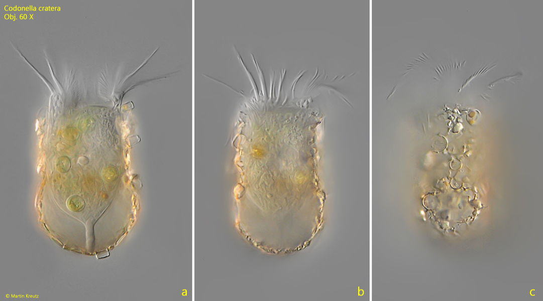
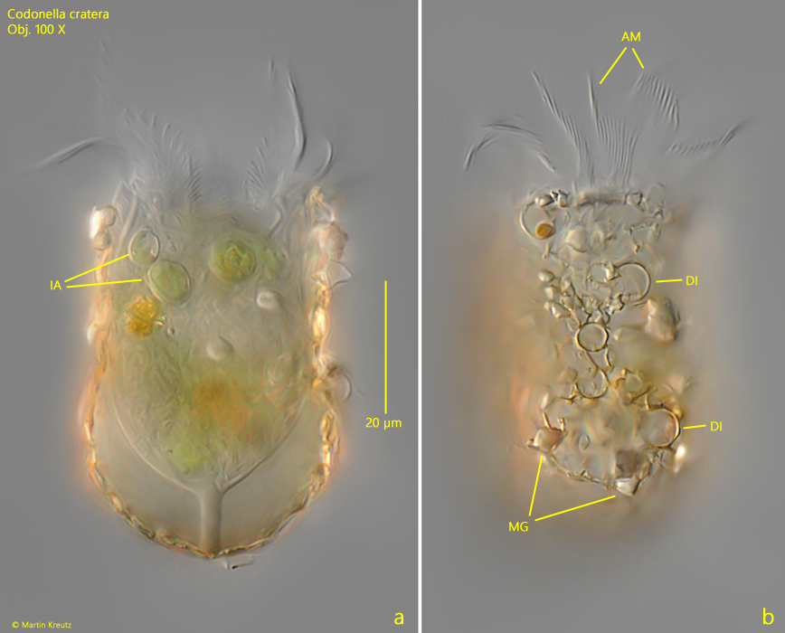
The ciliation of Codonella cratera is difficult to study as long as the ciliate is inside the shell. Essentially, it consists of about 30 rows of cilia, which run slightly spirally around the body. About 20 rows of these cilia are very close together and form a field of short cilia in the anterior third of the body.

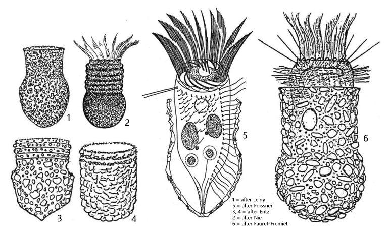
Due to the characteristic shape of the lorica, Codonella cratera cannot be confused with any other species.
Codonella-cratera

Fig. 1 a-c: Codonella cratera. L = 52 µm (of lorica). Three focal planes of a freely swimming specimen. Obj. 60 X.

Codonella-cratera

Fig. 2a-b: Codonella cratera. L = 52 µm (of lorica). The same specimen as shown in fig. 1 a-c in detail. AM = adoral membranelles, DI = diatoms, likely Cyclotella spec., IA = ingested algae cells, MG = mineral grains. Obj. 100 X.