connecting surfaces to neighboring cells flattened
one parietal chloroplast with one pyrenoid
planktonic lifestyle
Coelastrum astroideum
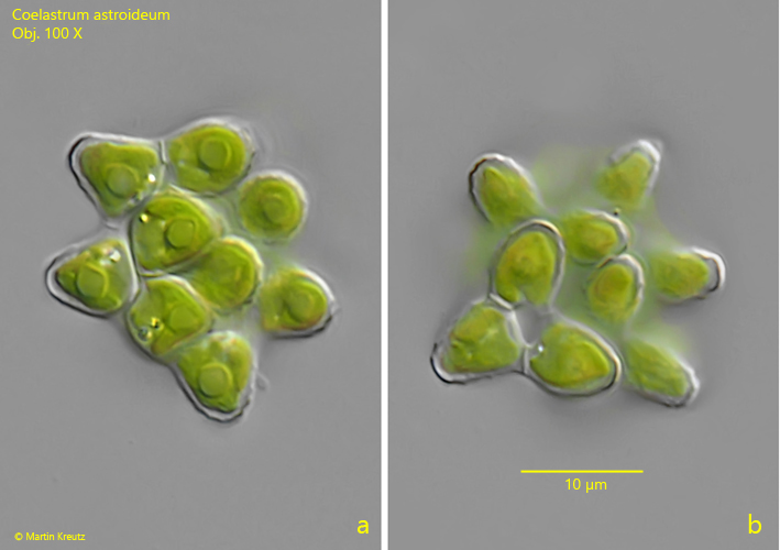
I found many coenobia of Coelastrum astroideum in the plankton of the pond of the waste disposal company Constance. The coenobia had a diameter of 20–30 µm and thus corresponded to the data given by Komarek & Trebon (1983). I found most coenobia in autumn.
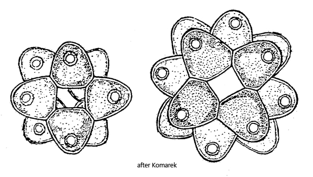
The cells in the coenobia were ovoid in shape as drawn by Komarek (s. drawing above). The tapered ends of the cells always points outward. The cell walls in my population were all slightly wrinkled (s. fig. 1 b).
Coelastrum astroideum differs from the similar species Coelastrum pseudomicroporum by the type of connection between the cells. In Coelastrum astroideum, there are only connections to the lateral neighboring cells, whereas in Coelastrum pseudomicroporum there are also connections at the basal surfaces of the cells to the inner cells.
Fig. 1 a-b: Coelastrum astroideum. D = 25 µm (of coenobium). A coenobium of 16 ovoid cells. The cell wall of the cells is slightly wrinkled. Obj. 100 X.