periplast smooth with subpellicular refractive granules
granules evert periplast in wart-like manner
sometimes with two terminal lobes
Collodictyon triciliatum
So far I have only found Collodictyon triciliatum sporadically in the plankton from the pond of the waste disposal company Constance. One has to examine the fresh plankton samples very carefully, because the slowly swimming specimens are easy to overlook.
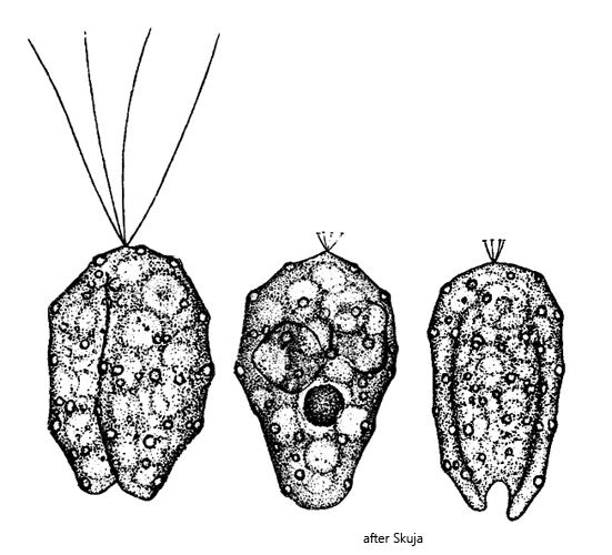
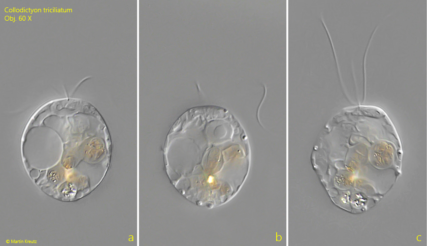
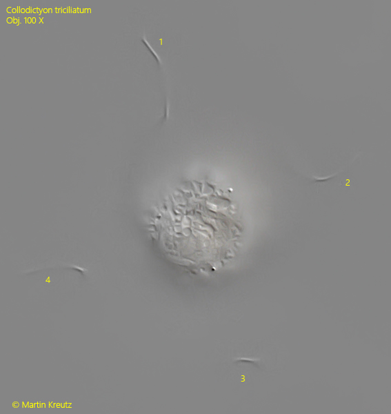
The name Collodictyon triciliatum, assigned by Carter (1865), is quite misleading, since this flagellate actually has 4 flagella of equal length. It was only Bütschli (1887) who recognized that Collodictyon triciliatum has 4 flagella. In 1919 a redescription of Collodictyon triciliatum by Rhodes appeared, in which the 4 flagella were confirmed. In lateral view, the number of flagella is difficult to determine. In fact, I was able to document them best in apical view (s. fig. 5).
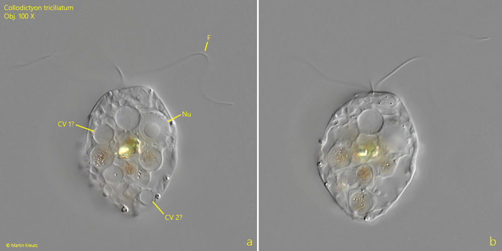
The cytoplasm of Collodictyon triciliatum is always strongly vacuolated and often also filled with many food vacuoles (s. fig. 3 a-b). As a result, the contractile vacuoles are difficult to detect. According to Skuja (1956) there are usually 1–2 contractile vacuoles. Rhodes (1919), on the other hand, was unable to reliably identify a contractile vacuole, and I too could not detect any actively functioning contractile vacuole.
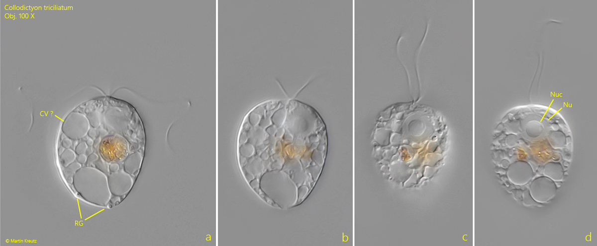
The nucleus is always located in the anterior third and has a large, central nucleolus (s. fig. 4 d). According to Skuja and Rhodes, Collodictyon triciliatum is supposed to possess a ventral groove that serves for food intake. Cytoplasmic pseudopodia can emerge from it, which enclose the prey organisms and lead them to phagocytosis. I was unable to observe this ventral groove in any of my specimens.
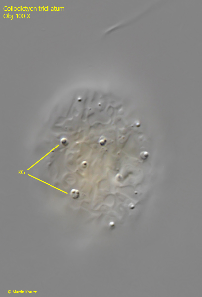
Beneath the periplast there are numerous highly refractive bodies (s. figs. 3 a and 4 a). These form wart-like elevations on the periplast, which gives the flagellates a characteristic appearance. Nothing is known about the nature of these granules. Based on my observations, they appear to have an oily character (s. fig. 6).
Fig. 1 a-c:Collodictyon triciliatum. L = 36 µm. An almost spherical freely swimming specimen. Obj. 60 X.
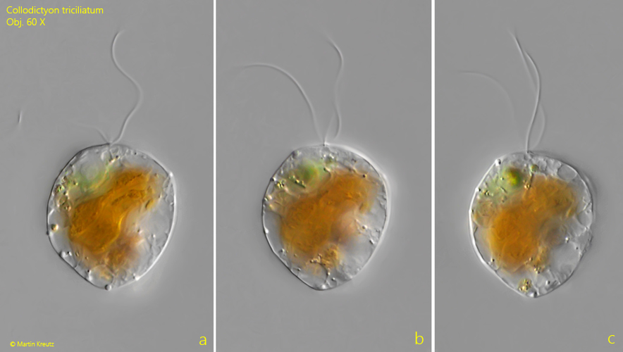
Fig. 2 a-c:Collodictyon triciliatum. L = 43 µm. A larger specimen with large phagocytosed prey. Obj. 60 X.
Fig. 3 a-b:Collodictyon triciliatum. L = 40 µm. An obovoid specimen. The nucleus (Nu) is located in the anterior third. Probably two contractile vacuoles are visible (CV 1, CV 2). F = flagella. Obj. 100 X.
Fig. 4 a-d:Collodictyon triciliatum. L = 34 µm. Different focal planes of a fourth freely swimming specimen. Note the spherical nucleolus (Nuc) in the nucleus (Nu). CV ? = probably one of the contractile vacuoles, RG = refractive granules. Obj. 100 X.
Fig. 5:Collodictyon triciliatum. A specimen in apikal view for visualization of the 4 flagella (1-4). Obj. 100 X.
Fig. 6:Collodictyon triciliatum. Focal plane on the subpellicular refractive granules. They appear to be similar to oil droplets. Obj. 100 X.