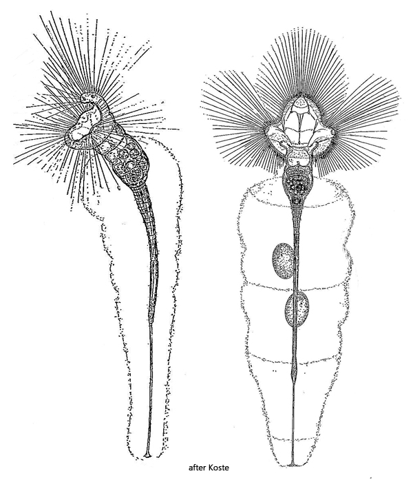
dorsal lobe the largest, lateral small, ventral medium sized
interspace between lobes with cilia
length up to 1400 µm
with long setae arise from the knobs
long, slender foot terminated by nonretractile peduncle
oval eggs deposited in gelatinous tube
in a gelatinuous tube
eyespots only in juvenile specimens
Collotheca campanulata
I regularly find Collotheca campanulata in the Simmelried, but usually only single specimens. However, in old samples, several specimens often settle on the vessel wall. Sometimes I also find specimens on the floating coverslip.
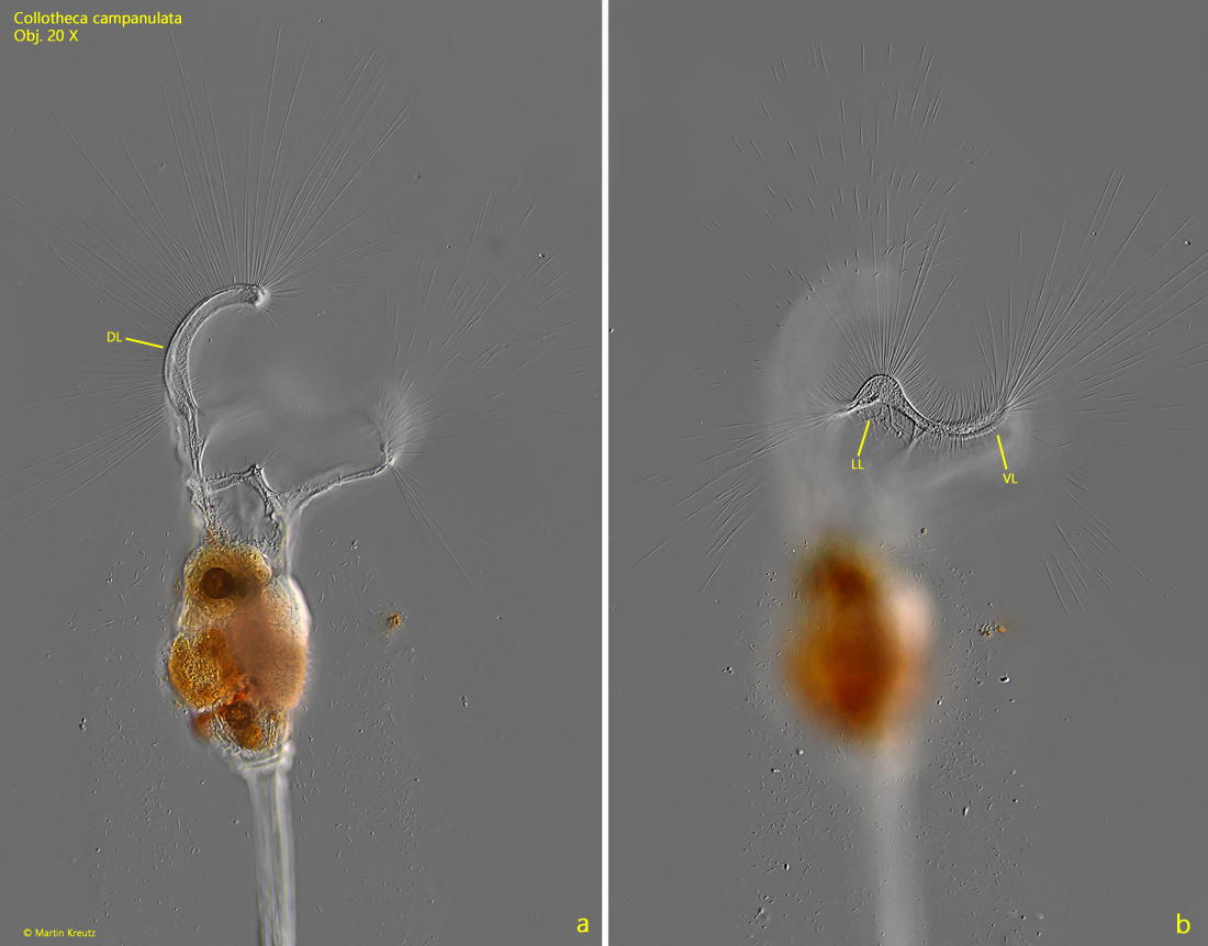
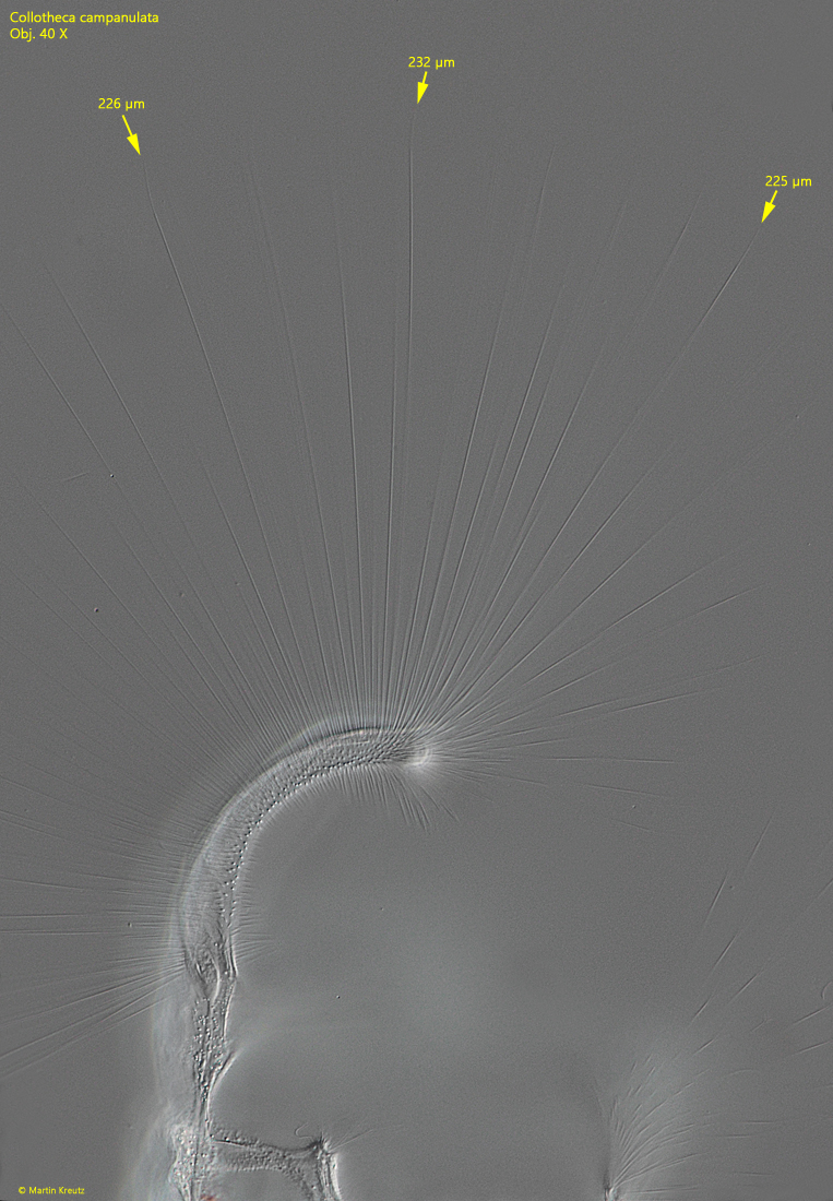
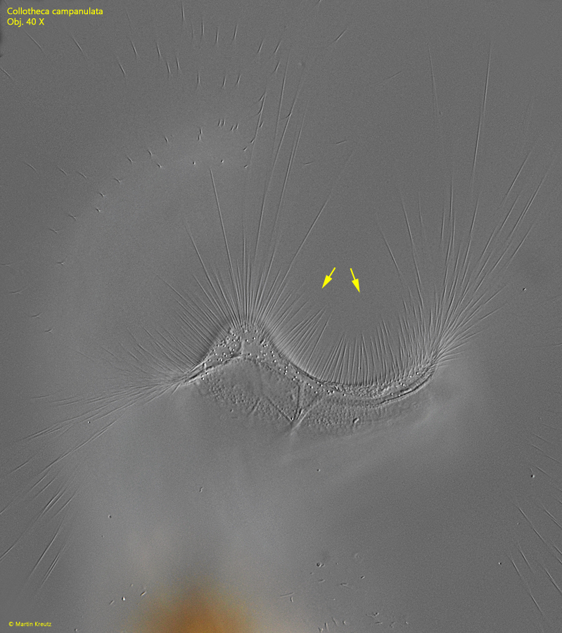
Collotheca campanulata can be recognized by the 5 clearly rounded lobes, which are not truncated but rounded (s. fig. 3 a-b). When the corona is fully extended, the dorsal lobe is the largest and distinctly bent inward. I was able to determine the length of the extended setae consistently at 220–230 µm (s. fig. 4), although this can vary between different individuals. The margin of the corona between the lobes is covered with shorter setae (s. fig. 5). This is also a distinguishing feature from the similar species Collotheca ornata, which is bare between the lobes.
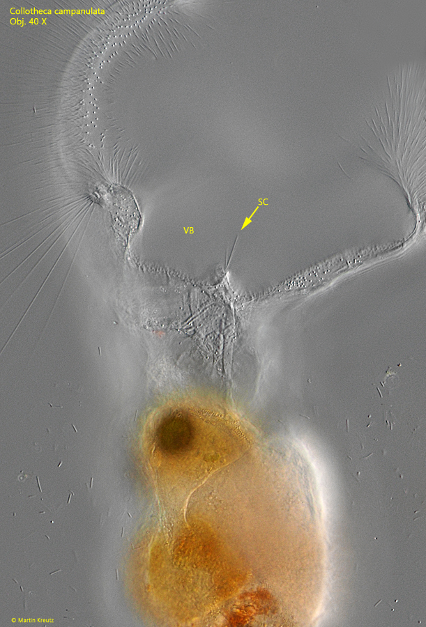
With the extended setae flagellates, algae, and small ciliates are caught, which then enter the vestibulum. At the bottom of the vestibulum, there are sensory cilia that detect the captured prey (s. fig. 6). Then the corona contracts briefly to transfer the prey into the underlying trochus with this movement. From here, the prey is then grabbed by the trophi, crushed, and transported into the crop.
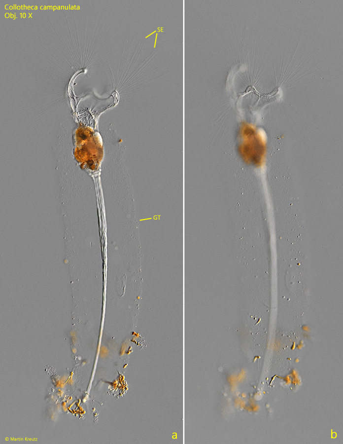
Fig. 1 a-b:Collotheca campanulata. L = 625 µm (without setae). Two focal planes of an elongated specimen in a gelatinous tube (GT). SE = setae. Obj. 10 X.
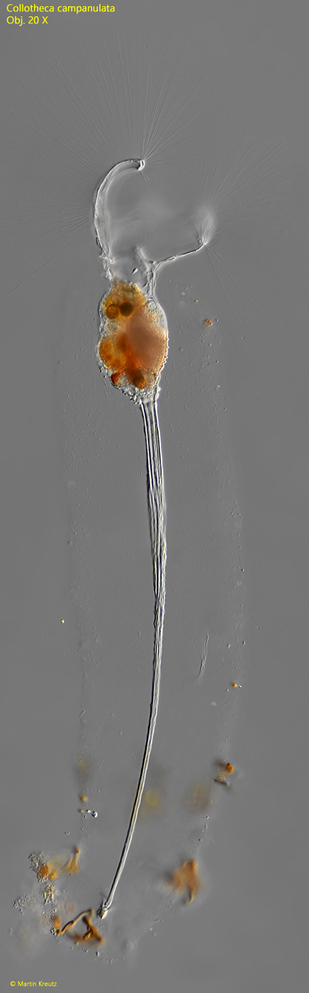
Fig. 2:Collotheca campanulata. L = 625 µm (without setae). The same specimen as shown in fig. 1 a-b at higher magnification. Obj. 20 X.
Fig. 3 a-b:Collotheca campanulata. Two focal planes of the corona. The dorsal lobe (DB) is bent inwards. The lateral lobes (LL) and the ventral lobe (VL) are small and rounded. Obj. 20 X.
Fig. 4:Collotheca campanulata. The setae of the dorsal lobe are contantly 220-230 µm long (arrows). The distal ends of the setae are also significantly tapered. Obj. 40 X.
Fig. 5:Collotheca campanulata. In the interspaces between the lobes shorter setae are present (arrows). Obj. 40 X.
Fig. 6:Collotheca campanulata. At the bottom of the vestibulum (VB) sensory cilia (SC) are located for detection of trapped prey. Obj. 40 X.