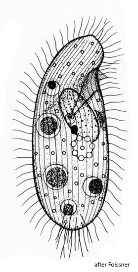
oral apparatus in anterior third, oriented obliquely
macronucleus spherical in center
one ellipsoid micronucleus
one contractile vacuole below cell equator
32–44 longitudinal rows of cilia
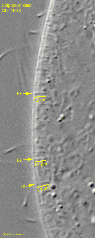
fringe of inconspicuous extrusomes
The specimens of Colpidium kleini shown below come from a culture that Holger Weis kindly made available to me. I myself have so far been unable to detect Colpidium kleini at my local sampling sites, although the species is described as very common.
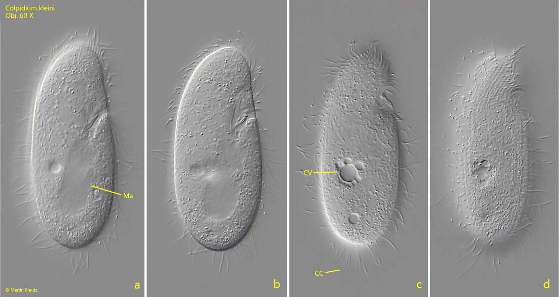
Colpidium kleini is characterized by a slender, somewhat kidney-shaped body. The anterior third is slightly slendered and inclined somewhat ventrally. The specimens were all between 70–75 µm long. The mouth opening is located in the anterior third. At the posterior end there are some elongated caudal cilia (s. fig. 1 c).
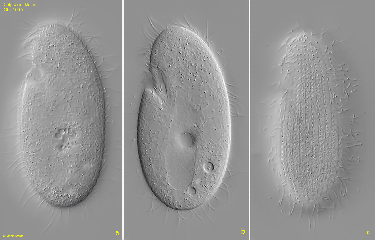
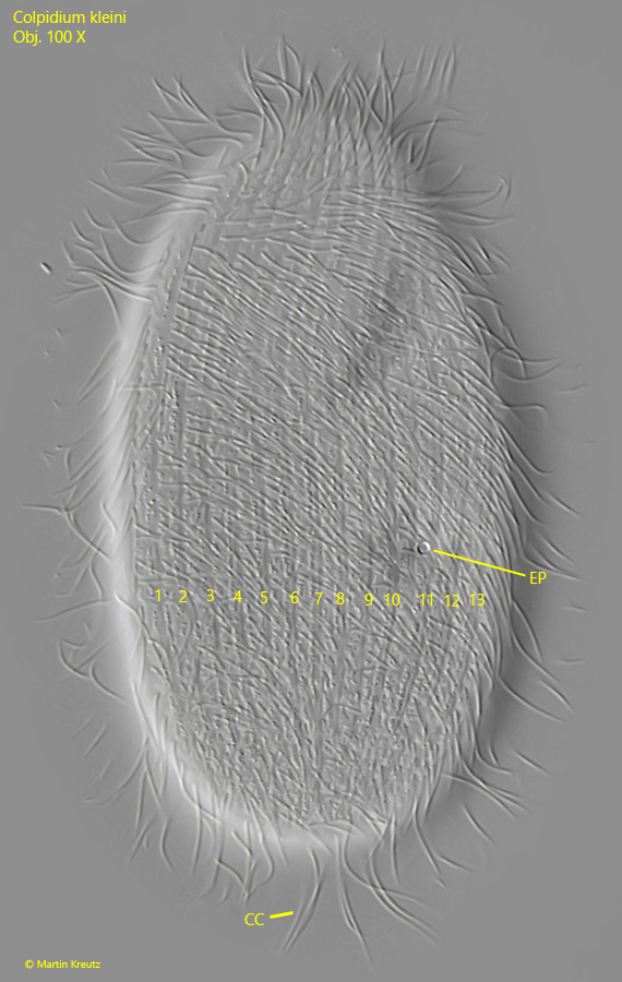
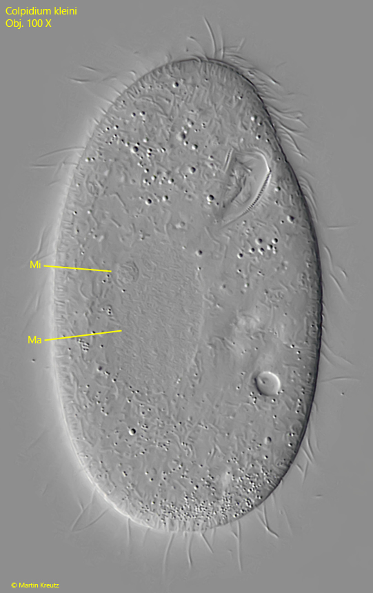
An important distinguishing feature from the similar species Colpidium colpoda is the number of longitudinal rows of cilia. In a slightly compressed specimen, I was able to count 13 rows (s. fig. 4). Calculated over the entire circumference, there are about 30–36 rows. This makes it possible to rule out Colpidium colpoda (50–63 rows). For Colpidium kleini 32–44 longitidinal rows of cilia are specified what fit my observations. The second similar species Dexiostoma campylum can also be ruled out by the position of the contractile vacuole (in posterior third) and the shape of the macronucleus (spherical). In my specimens the contractile vacuole was located shortly below the cell equator (s. fig. 1 c) und in all specimens the macronucleus was ellipsoid (s. fig. 5). I attribute the small size of my specimens (70–75 µm) on the culture conditions. The common size for Colpidium kleini is about 100 µm.
Fig. 1 a-d:Colpidium kleini. L = 72 µm. A freely swimming specimen from right. CC = caudal cilia, CV = contractile vacuole, Ma = macronucleus. Obj. 60 X.
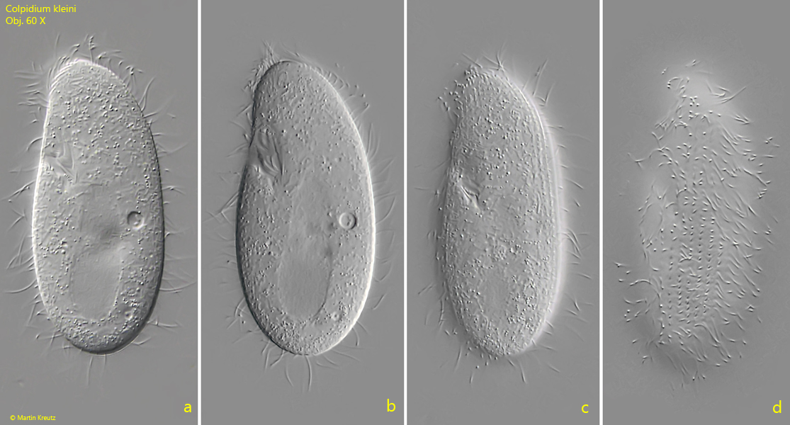
Fig. 2 a-d:Colpidium kleini. L = 72 µm. A second freely swimming specimen from left. Obj. 60 X.
Fig. 3 a-c:Colpidium kleini. L = 72 µm. The same specimen as shown in fig. 2 a-d at higher magnification. Obj. 60 X.
Fig. 4:Colpidium kleini. Focal plane on the excretion pore (EP) and longitudinal rows of cilia in a slightly squashed specimen. In this view (dorso-lateral from right) 13 longitudinal rows of cilia (1–13) are visible. CC = caudal cilia. Obj. 100 X.
Fig. 5:Colpidium kleini. The ellipsoid macronucleus (Ma) and the spherical micronucleus (Mi) in a slightly squashed specimen. Obj. 100 X.
Fig. 6:Colpidium kleini. The delicate, rod-shaped extrusomes (EX) beneath the pellicle are 1.4–1.7 µm long. Obj. 100 X.