Crucigenia mucronata

(G.M. Smith) Komárek, 1974

Most likely ID: n.a.


Synonym: n.a.


Sampling location: Pond of the waste disposal company Constance


Phylogenetic tree: Crucigenia mucronata


Diagnosis: 

  • coenobia square with square gap in center
  • forming composite syncoenobia without gelatinous sheath
  • cells irregular oval or trapezoid
  • outer side of cell almost straight, inner side convex
  • apices slightly tapered with warts
  • length 6–9 µm, width 3–6 µm
  • chloroplast parietal, one pyrenoid
  • planktonic lifestyle
Crucigenia-mucronata
Crucigenia mucronata
So far, I have found Crucigenia mucronata only once in the plankton of the pond of the waste disposal company Constance. This pond is highly eutrophic. This matches the descriptions by Komarek & Fott (1983), who described it as a rare species in the plankton of eutrophic waters.
 
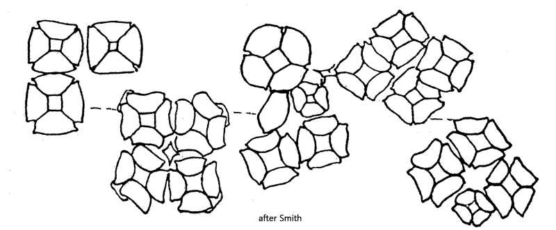
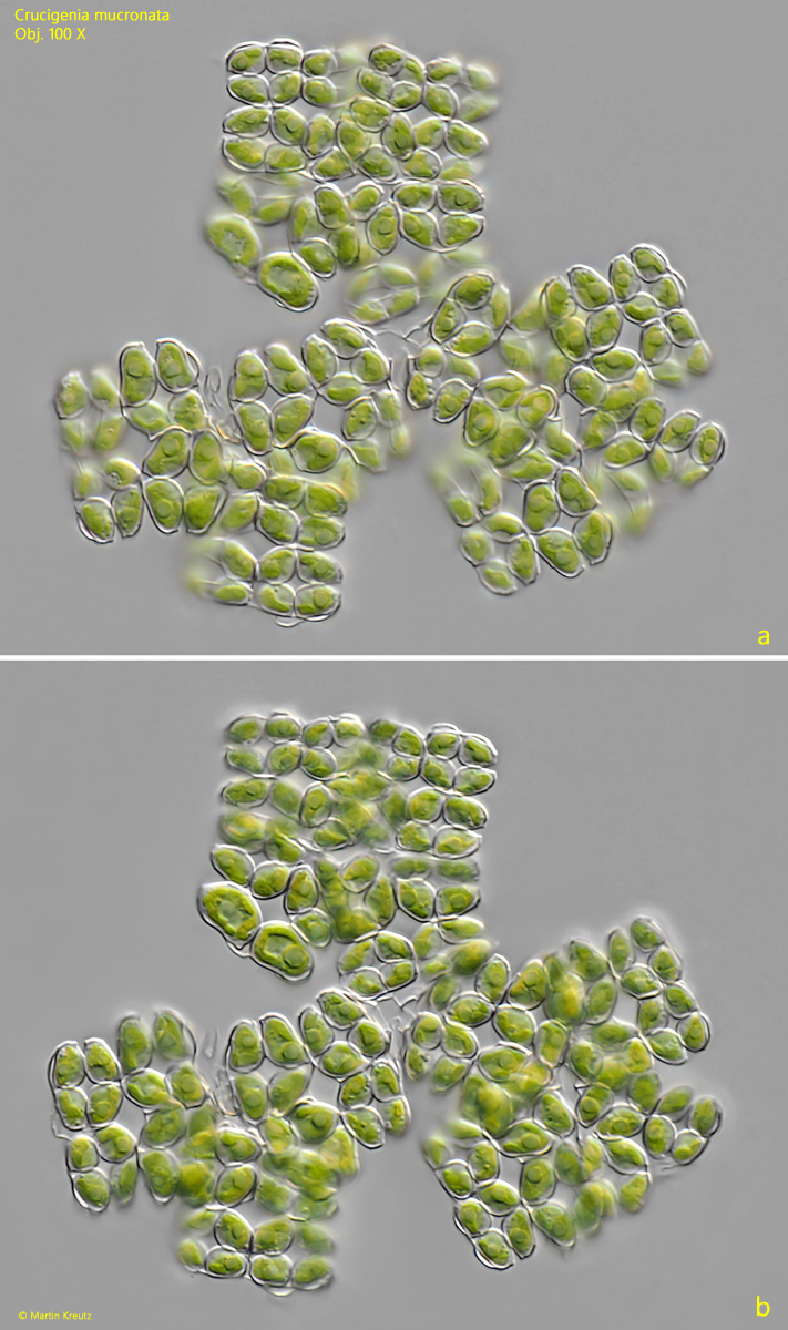
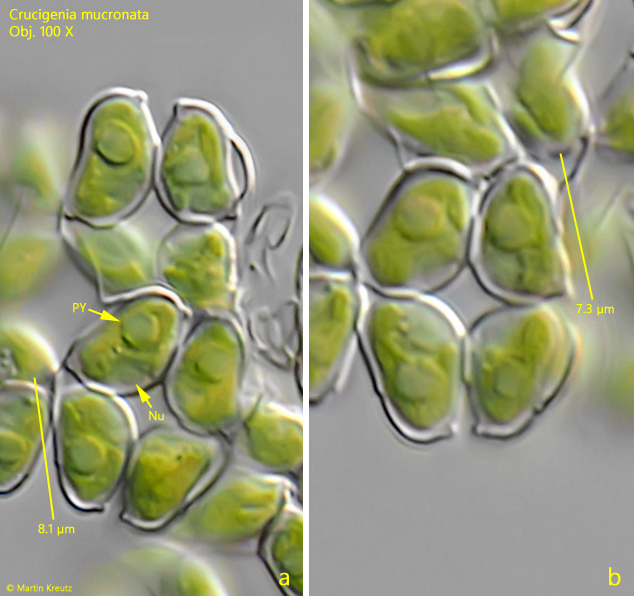
In my finding, it was a large, contiguous synzoenobium consisting of about 10–15 coenobia with 8–16 cells. In the coenobia, the cells were arranged in a square pattern. The cells were either flat or slightly convex on the outward-facing side, while the inward-facing side was distinctly convex (s. fig. 2 a-b). The cells had a length of 7.0–8.5 µm. The apices were shaped into short warts, as is typical for this species (s. fig. 2 a-b). The pyrenoid was clearly visible, as was the cell nucleus in the center of the cell.
Crucigenia-mucronata

Fig. 1 a-b: Crucigenia mucronata. D = 90 µm (of syncoenobium). Two focal planes of a syncoenobium of several coenobia of 8-16 cells. Obj. 100 X.

Crucigenia-mucronata

Fig. 2 a-b: Crucigenia mucronata. L = 7.0–8.5 µm (of cells). Two crops of the fig. 1 a-b with the cells in detail. Note the wart-shaped protuberances at the apices of the cells. Nu = nucleus, PY = pyrenoid. Obj. 100 X.