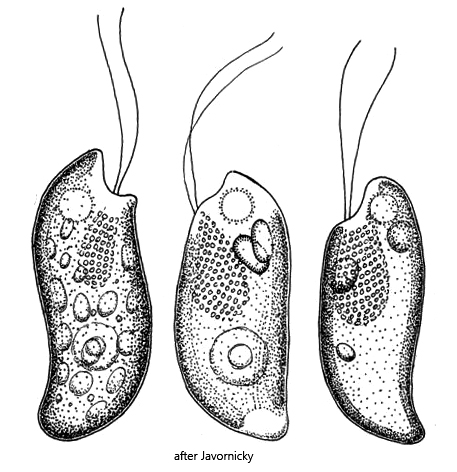
cells spindle-shaped, posterior end curved dorsally
posterior end pointed
cells sometimes slightly sigmoid
laterally flattened
length 20–46 µm
gullet reach almost cell equator
two Maupas’ bodies, sometimes absent
two inconspicuous pyrenoids
two chromatophores, olive-green or reddish-brown
two flagella
contractile vacuole apical
nucleus in posterior third
Cryptomonas reflexa var. reflexa
I found Cryptomonas reflexa var. reflexa in the Simmelried in October 2026. At this point, the Simmelried had almost completely dried out after a months-long dry period. The sample comes from a small puddle that remained as a remnant of a pond. The specimens of Cryptomonas reflexa var. reflexa accumulated on the surface of the sample container.
The taxonomy of cryptomonads is undergoing major changes and in many respects has not been conclusively clarified. I carried out the identification of Cryptomonas reflexa var. reflexa essentially using the publication by Javornicky (2014). This publication seems to me to reflect the current state of taxonomy.
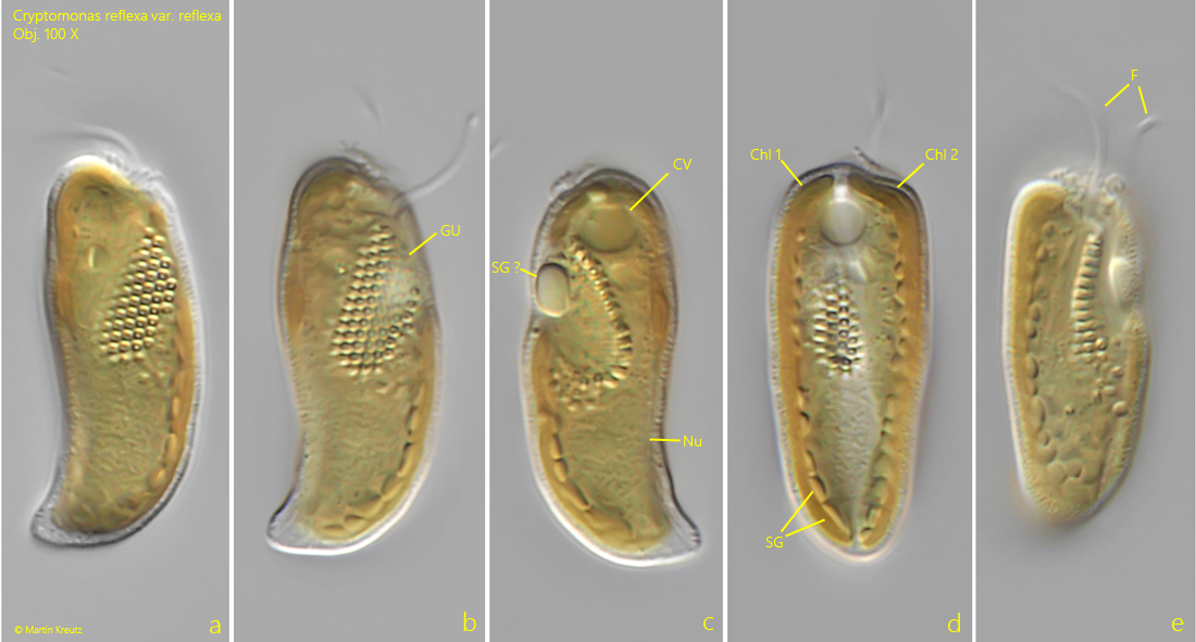
Cryptomonas reflexa var. reflexa has a characteristic shape. The body tapers evenly toward the posterior end and is pointed. The pointed posterior end is clearly curved backward. The gullet, which is lined with numerous ejectisomes, begins in an apical depression and extends almost to the middle of the body. Laterally two chloroplasts are present. According to Javornicky they are supposed to have a connection on the dorsal side. Unfortunately, I was not able to detect this connection.
The specimens of my populations did not possess Maupas’ bodies. I was unable to discern the pyrenoids (there are supposed to be two of them), although Javornicky mentions that they can be very inconspicuous. The length of my specimens ranged between 30–36 µm.
Fig. 1 a-e:Cryptomonas reflexa var. reflexa. L = 34 µm. A freely swimming specimen from right (a, b), left (c) and from ventral (d, e). Chl 1 + 2 = chloroplasts, CV = contractile vacuole, F = flagella, GU = gullet, Nu = nucleus, SG = starch grains, SG ? = probably a starch grain. Obj. 100 X.