So far, I have only found two specimens of Cyclidium flagellatum. I found both specimens among decaying masses of plants in August and October 2021 in the Simmelried.
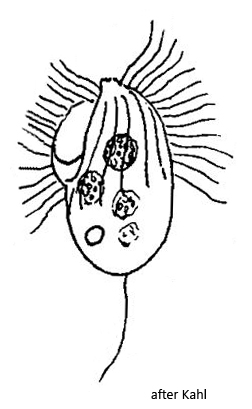
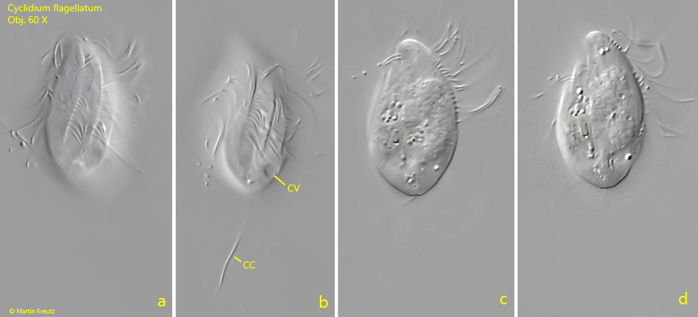
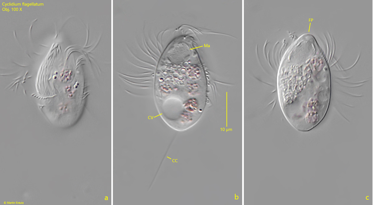
In the fresh samples, Cyclidium flagellatum stands out due to its rowing-like swimming movement. The somatic cilia are very long and soft. According to my measurements, they are 12 µm long, which corresponds to almost half the body length. I was able to recognize all the features as described by Kahl. The frontal plate is snout-shaped. The somatic cilia run in slightly spiral rows clockwise up to the posterior third of the body (s. fig. 1 a-b). The posterior third of the body is naked. The food vacuoles mainly contained purple bacteria. The macronucleus was located near the anterior end and the contractile vacuole is clearly subterminal (s. fig. 2 b). No extrusomes were visible. The caudal cilium was about one body length long. With a length of 28 µm, my specimens were exactly in the middle of the range given by Kahl.
Fig. 1 a-d:Cyclidium flagellatum. L = 28 µm. A freely swimming specimen. Note the longitudinal rows of cilia running clockwise slightly spirally (a, b). The posterior third is naked. CC = caudal cilium, CV = contractile vacuole. Obj. 60 X.
Fig. 2 a-c:Cyclidium flagellatum. L = 28 µm. A second freely swimming specimen. The macronucleus (Ma) is located in the anterior end below the snout-shaped frontal plate (FP). The contractile vacuole (CV) is located in the posterior fifth. CC = caudal cilium. Obj. 100 X.