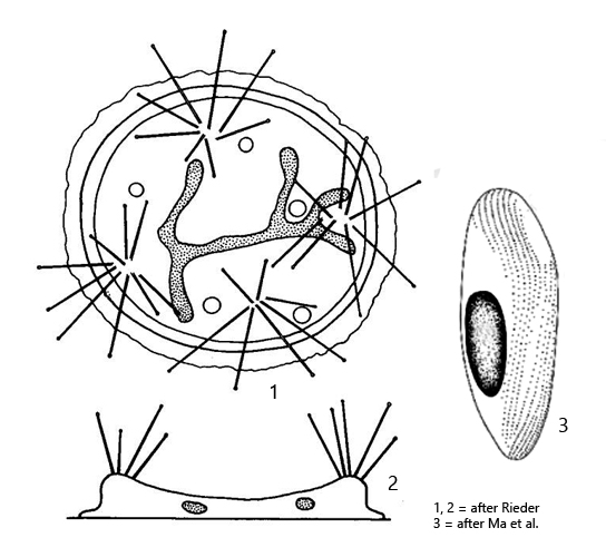
macronucleus ribbon-shaped, branched or horseshoe-shaped
3–6 bundles of tentacles (rarely up to 14)
tentacles up to 500 µm long
4–8 contractile vacuoles in periphery
swarmer ellipsoid, about 100 µm long
Cyclophrya magna
I found Cyclophrya magna on the stems of water lilies in the pond of the waste disposal company Constance. Adhering to the stems, the specimens are difficult to observe. Therefore, I placed some cut stems with some water from the site into a petri dish and placed a few coverslips on the water surface. After a few days, the first specimens had settled on the floating coverslips and could then be easily observed.
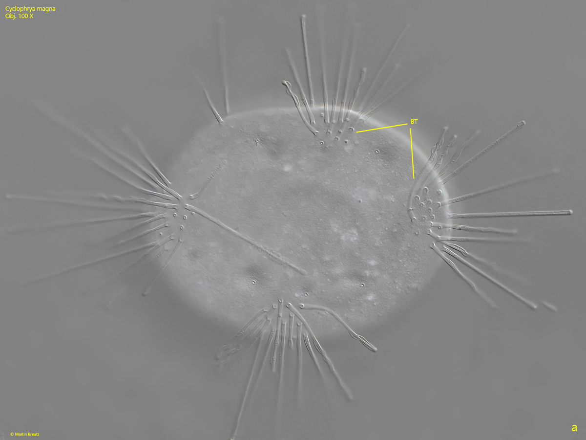
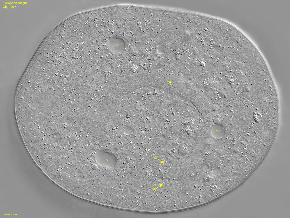
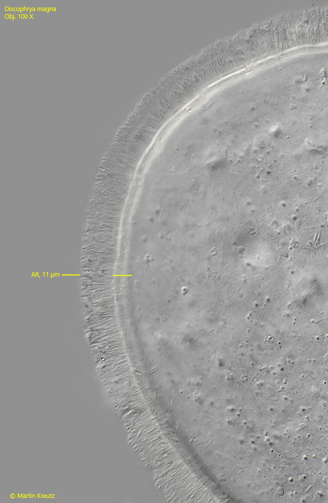
The specimens of Cyclophrya magna in my population had a diameter of 160–185 µm and were slightly elliptical in shape. The macronucleus was horseshoe-shaped in all specimens with at most one branch. Matthes (1988) does not provide any information on the number of micronuclei. I could usually only recognize 2–3, but there are probably more (s. fig. 3). Most specimens had 4–6 contractile vacuoles, and 4–5 bundles of tentacles. Each bundle consisted of 10-17 tentacles (s. fig. 2 a). A characteristic feature of Cyclophrya magna is the adhesive rim, with which the suctor holds itself onto the substrate. It has a distinct radial striation and was usually 11–12 µm wide in my specimens (s. fig. 6). I could not observe a swarmer or the formation of a swarmer. According to the observations of Ma et al. (2021), it is ellipsoid in shape, about 100 µm long, without tentacles and has an ellipsoid macronucleus (s. drawing 3, above).
Cyclophrya differs from the similar genus Heliophrya in size, the shape of the macronucleus, and the structure of the adhesive rim. All species of the genus Heliophrya are smaller than 100 µm, the macronucleus is globular or kidney-shaped, and the adhesive rim is homogeneous without radial striation.
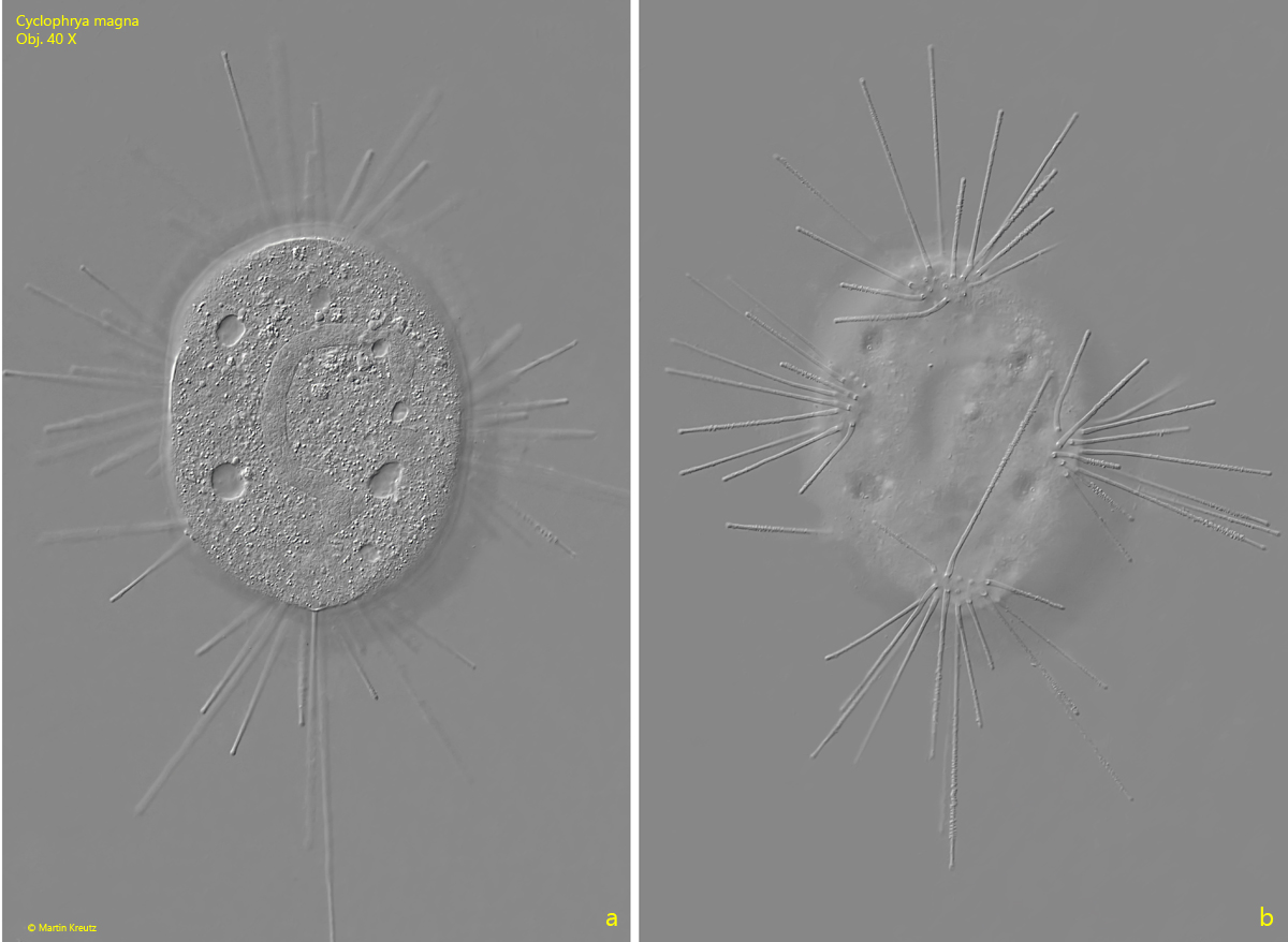
Fig. 1 a-b:Cyclophrya magna. D = 183 µm. Two focal planes of a specimen attached to a floating coverslip. Obj. 40 X.
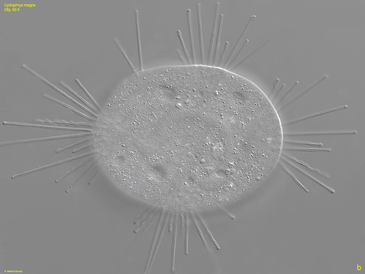
Fig. 2 a-b:Cyclophrya magna. D = 183 µm. Two focal planes on the bundles of tentacles (BT). Obj. 60 X.
Fig. 3:Cyclophrya magna. Focal plane on the horseshoe-shaped macronucleus (Ma) in a slightly squashed specimen. Note the two visible micronuclei (Mi) adjacent to the macronucleus. CV = contractile vacuoles. Obj. 100 X.
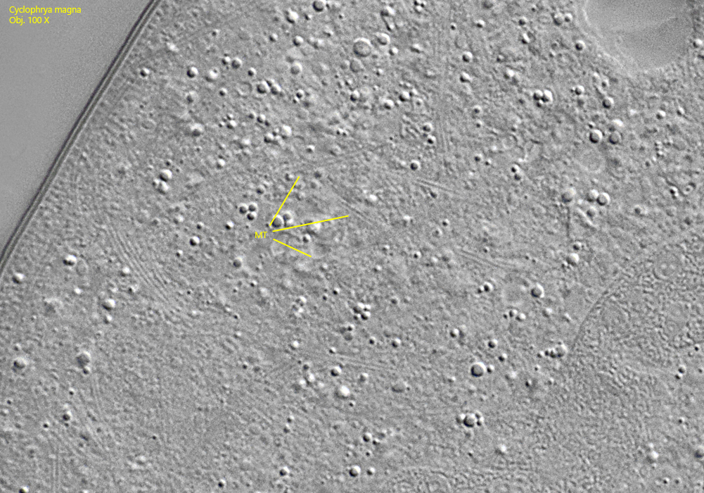
Fig. 4:Cyclophrya magna. Near the resolution limit, fragments of microtubules (MT) can be seen in the cytoplasm, which are presumably kept in reserve for the construction of the tentacles or result from their degradation. Obj. 100 X.
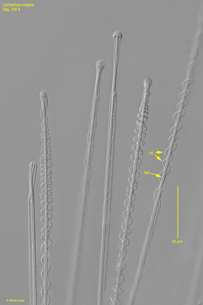
Fig. 5:Cyclophrya magna. The tentacles in detail. The interior of the tentacle consists of a rod made up of microtubules (MT). This is surrounded by the pellicle (PE) of the suctor. The pellicle is very clearly visible when it folds as the tentacle retracts. Obj. 100 X.
Fig. 6:Cyclophrya magna. The suctor is attached to the substrate with an adhesive rim (AR). The rim is clearly striated and has in this specimen a width of 11 µm. Obj. 100 X.