So far, I have only found Desmidium grevillei in the Schwemm Moor in Austria. The algae was very common in the samples.
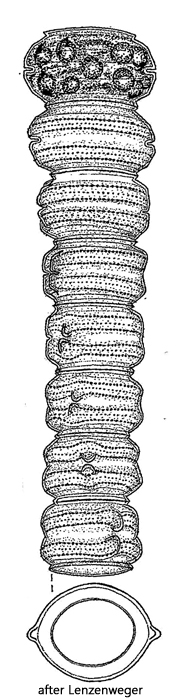
The cells in the filaments of Desmidium grevillei consist of two semi-cells, like many other desmid algae (e.g. Cosmarium or Staurastrum). However, the isthmus is only very weakly pronounced and is only a slight tapering. Each semi-cell has conical basal angles. Here, the cell wall is also significantly thickened. Since the cells in the filament are always slightly offset from each other, these conical thickenings appear to run spirally around the filament (s. fig. 4). Each semi-cell also has a ring of fine pores, which are only visible under high magnification (s. fig. 5).
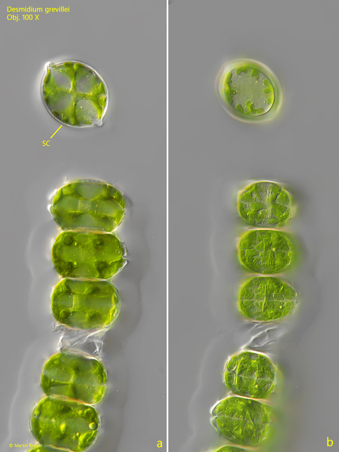
In lateral view, the individual cells appear almost rectangular. They are connected to each other with their apices. If a cell is detached from the filament and rotated, it can be viewed in apical view (s. fig. 3 a-b). The shape is similar to that of a lemon. The conical ends are the basal angles of the semi-cells.
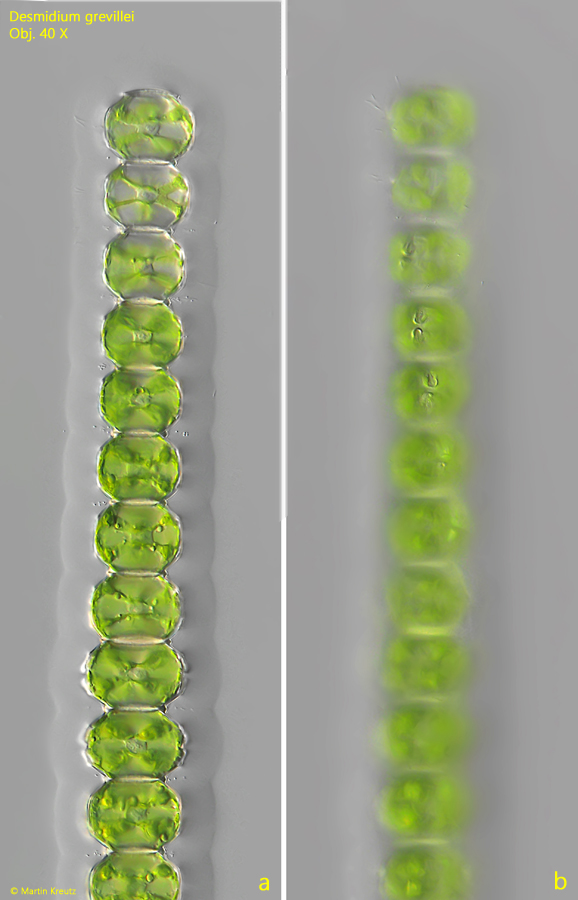
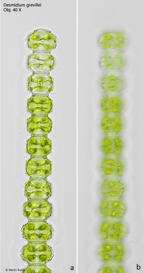
Fig. 1 a-b:Desmidium grevillei. L = 30–32 µm (of cells). Two focal planes of the end of a filament in DIC. Obj. 40 X.
Fig. 2 a-b:Desmidium grevillei. L = 30–32 µm (of cells). The same specimen as shown in fig. 1 a-b in brightfield illumination. Obj. 40 X.
Fig. 3 a-b:Desmidium grevillei. Two focal planes of a slightly squashed end of a filemant with a sepated cell (SC). The separate cell is visible in apical view with an elliptical shape and conical shaped basal angles. Obj. 100 X.
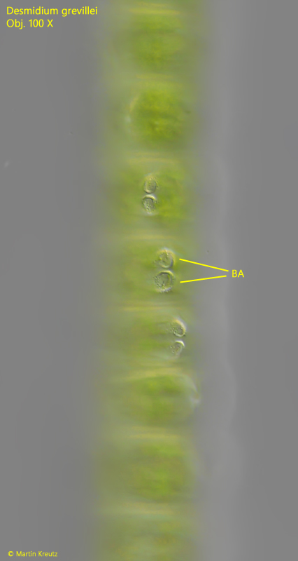
Fig. 4:Desmidium grevillei. The cells in the filament are arranged with an offset to each other. The focal planes is on the conical shaped basal angles (BA) of the cells. Obj. 40 X.
Fig. 5:Desmidium grevillei. The cell wall of the semi-cells are covered with a ring of granules (GR). Obj. 100 X.