unbranched filaments of triangular (rarely quadrangular) cells
filaments are twisted, covered by mucilage
cells 35–50 µm wide, 12–21 µm long
cells constricted in the middle
each apical angle of cells with short process for connection
chloroplast of each semi-cell with 3 pyrenoids
Desmidium swartzii
Desmidium swartzii is the most common filamentous desmid and is not only found in bogs. In my sampling site Simmelried it is very common and can usually be found between floating plant masses.
The filaments of Desmidium swartzii are not branched and can be easily recognized even at low magnifications, because the trigonal cells are connected slightly offset to each other and thus the filament appears twisted (s. figs. 1 a-b and 2 a-c). The cells themselves consist of two half-cells, which are, however, strongly widened and flattened. Like the cells of the genus Staurastrum, they have an isthmus in the middle (s. fig. 3 a-b). Each half-cell has a chloroplast with three pyrenoids. At the outer angles of the cells there are contact surfaces with which the cells are connected to the neighboring cells (s. fig. 4). When the coverslip pressure is increased, these connections break and the trigonal shape of the cells can be seen in apical view (s. fig. 5).
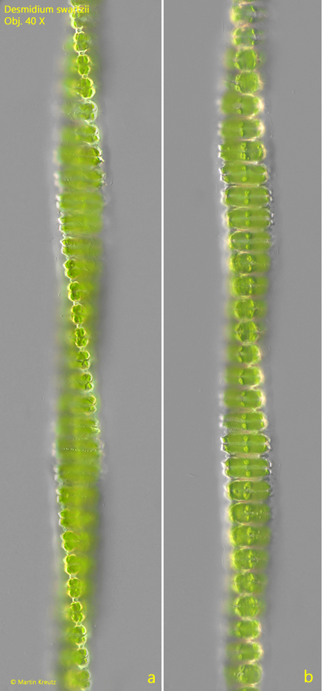
Fig. 1 a-b:Desmidium swartzii. Two focal planes of a twisted filament. Obj. 40 X..
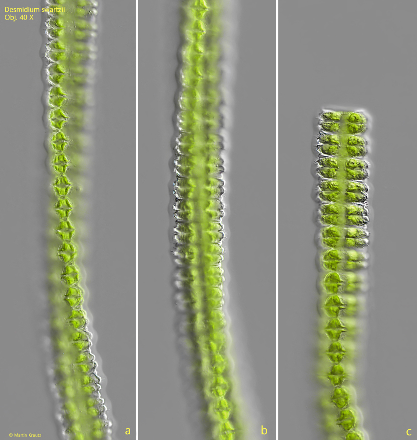
Fig. 2 a-c:Desmidium swartzii. L = 19–21 µm (of cells). Two focal planes of a filament of cells (a, b) and the end of the filament (c). Note the twisted arrangement of the cells with the filament. Obj. 40 X.
Fig. 3 a-b:Desmidium swartzii. L = 19–21 µm (of cells). The cells (CE) of the filament in detail. Near the isthmus of the cells rows of granules (GR) are visible. In each semi-cell (SC 1, SC 2) two of the three pyrenoids (PY) are visible. Obj. 100 X.
Fig. 4:Desmidium swartzii. Focal plane on the apical angle of the cells in the filament. Each semi-cell (SC 1, SC 2) is connected with the neighboring cells (arrows). Obj. 100 X.
Fig. 5:Desmidium swartzii. Under pressure of the coverslip the filaments desintegrates and the triangular shape of the cells in apical view become visible. Obj. 40 X.