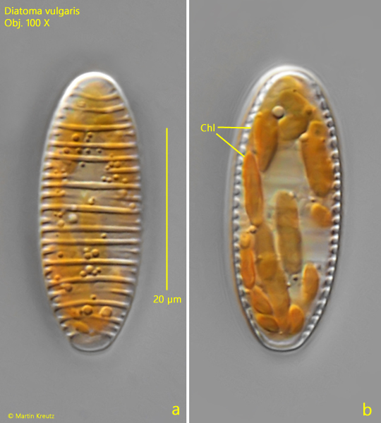
in valve view ellipsoid with slightly irregular transverse striae
several elongated ellipsoid chloroplasts
chloroplass golden brown or yellowish
Diatoma vulgaris
Diatoma vulgaris is a very common diatom, which can also be found in flowing waters. I mainly find it on the shore of Lake Constance as dark brown or orange-brown growth on stones and in the overflow of the Mühlhalden pond.
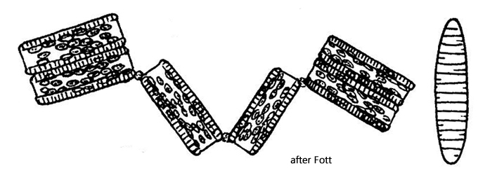
Diatoma vulgaris forms typical zig-zag bands of usually a maximum of 20 cells (s. fig. 1 a-b). The cells are connected at the diagonal corners of each cell by a gelatinous mass (s. fig. 2 a). The cells contain many chloroplasts, which are elongated ellipsoids and lie inside the shell. In girdle view, the cells have a rectangular shape. As the only structure, several parallel-running intercalary bands can be seen in this view (s. fig. 2 c). In valve view, the cells have an ellipsoid shape and characteristic transverse striation with large intervals (s. figs 3 a-c and 4 a-b). The striae have a slightly irregular shape.
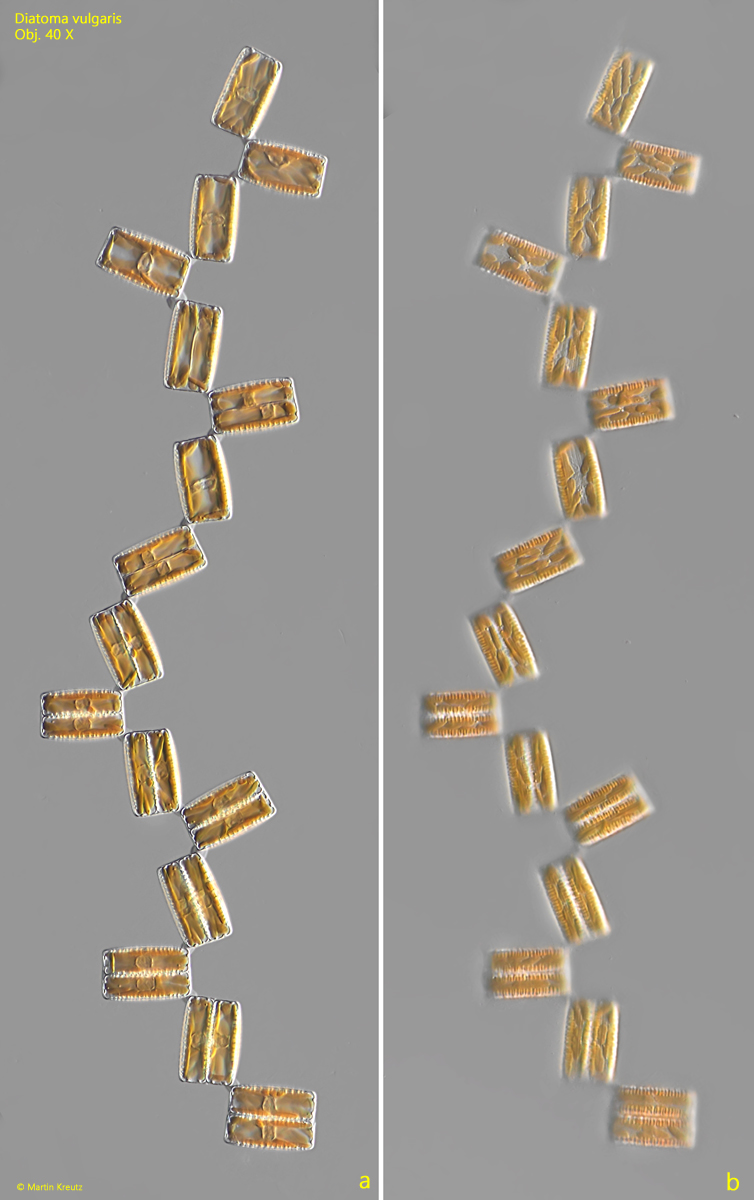
Fig. 1 a-b:Diatoma vulgaris. L = 32–37 µm (of cells). Two focal planes of a zig-zag chain of 16 cells in girdle view. Obj. 40 X.
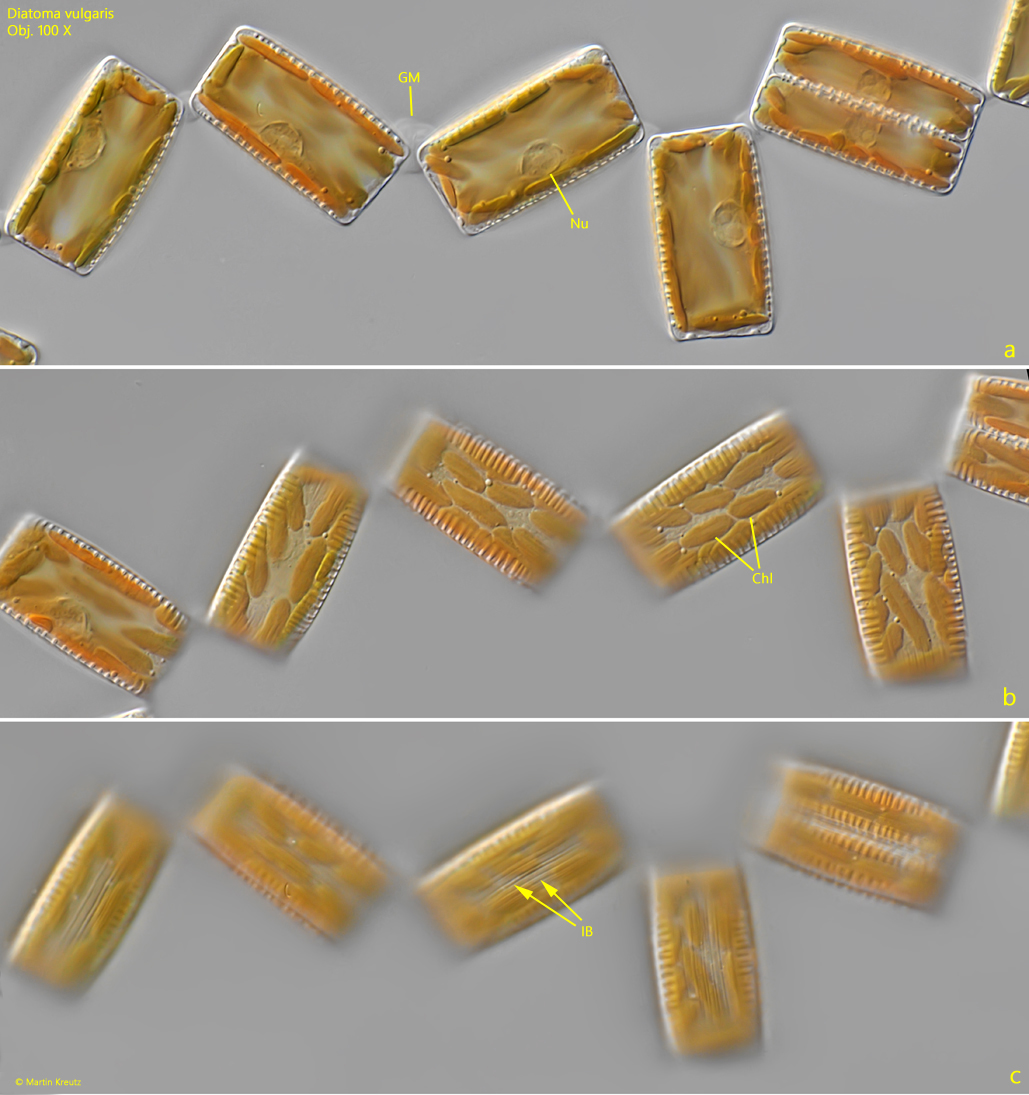
Fig. 2 a-c:Diatoma vulgaris. L = 35–38 µm (of cells). Three focal planes of some cells in girdle view. The cells are connected via a gelatinous mass (GM) at the corners of the cells. Chl = chloroplasts, IB = intercalary bands, Nu = nucleus. Obj. 100 X.
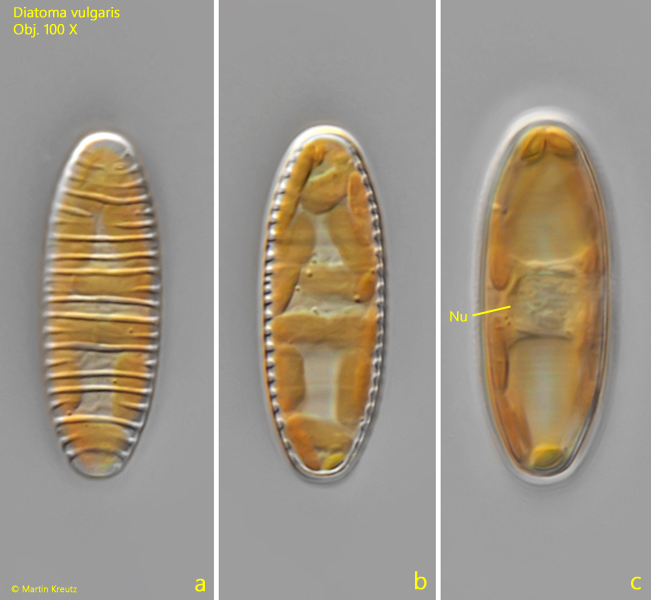
Fig. 3 a-c:Diatoma vulgaris. L = 38 µm. Three focal planes of a cell in valve view. The valve has an ornamentation of slightly irregular transverse striae. Nu = nucleus. Obj. 100 X..
Fig. 4 a-b:Diatoma vulgaris. L = 35 µm. A second cell in valve view. Chl = chloroplasts. Obj. 100 X.