I find Dimorphococcus lunatus very frequently in the plankton samples from various locations. This alga is particularly common in the heavily eutrophic pond of the waste disposal company Constance.
The colonies are covered in a gelatinous envelope and have usually a diameter of 50–100 µm. However, they do not consist of an accumulation of individual cells, but of coenobia of 4 cells each, which are located at the end of gelatinous stalks. This is why the colonies are called syncoenobia. The cells in each coenobium are present in two forms. The younger, inner cells are almost ellipsoidal, while the outer cells are kidney-shaped (s. fig. 4). When the syncoenobia are squashed under the coverslip, they disintegrate into the 4-cell coenobia.
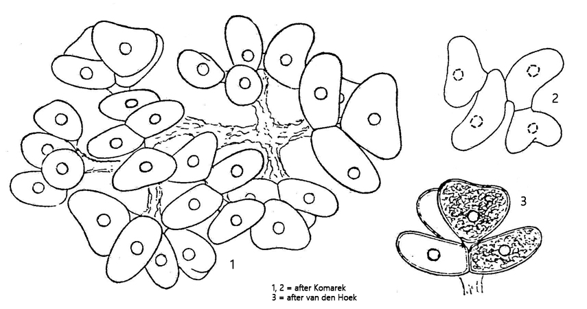
Fig. 1:Dimorphococcus lunatus. D = 30–60 µm (of syncoenobia). An accumulation of some syncoenobia in a plankton sample. Obj. 20 X.
Fig. 2:Dimorphococcus lunatus. D = 50–70 µm (of syncoenobia). Three slightly squashed syncoenobia. Obj. 40 X.
Fig. 3 a-b:Dimorphococcus lunatus. L = 11–15 µm (of cells). Two focal planes of a slightly squashed syncoenobium. Note the young elliptical cells and the older kidney-shaped cells. Obj. 100 X.
Fig. 4:Dimorphococcus lunatus. L = 17–19 µm (of cells). A squashed coenobium of 4 cells. The inner cells are ellipsoid while the outer cells are kidney-shaped. Nu = nucleus, PY = pyrenoid. Obj. 100 X.