So far, I have only found Dinobryon bavaricum twice. In June 2000 in the plankton of Lake Constance and 25 years later in July 2025 in the plankton of the Mühlweiher Litzelstetten. In both cases, I only found very small colonies of no more than three individuals.
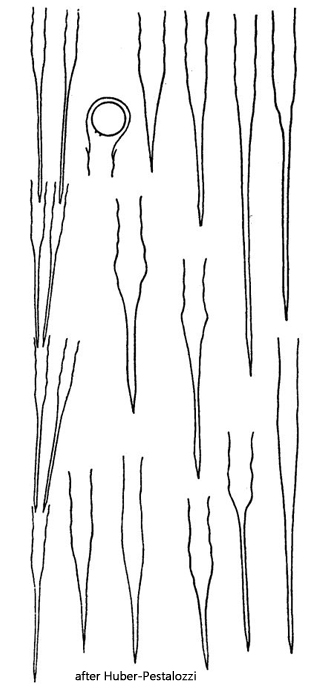
The most important distinguishing feature of Dinobryon bavaricum is the wavy walls of the loricae (s. fig. 1 a). This waveform can vary in intensity, but is always present. The loricae are also very slender and elongated. The aperture of the lorica is shaped cylindrically and only slightly widened, never funnel-shaped.
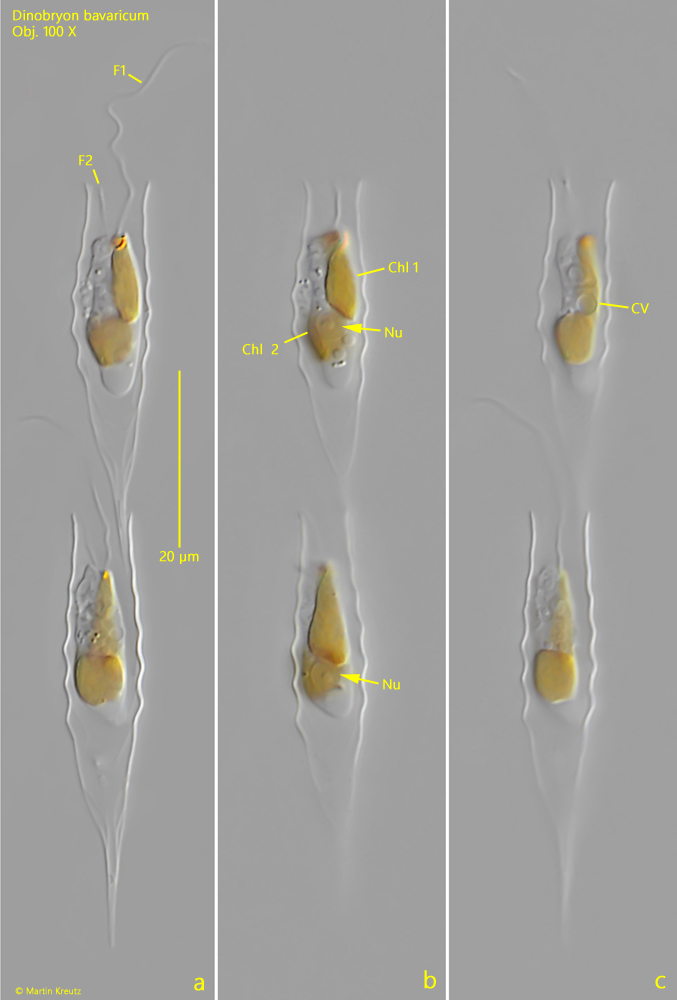
The loricae of my population were only 45–50 µm long, which is at the lower end of the range of 50–120 µm given by Huber-Pestalozzi (1941). The internal structure of the cells, with two chloroplasts, a centrally located cell nucleus, and a contractile vacuole, corresponded to the Dinobron type.
Fig. 1 a-c:Dinobryon bavaricum. L = 45–47 µm (of lorica). Three focal planes of two freely swimming specimens. Note the undulate walls of the loricae. Chl 1 + Chl 2 = chloroplasts, CV = contractile vacuole, F1 + F2 = flagella of unequal length, Nu = nucleus. Obj. 100 X.