I find Diplophrys archeri frequently and regularly in the Simmelried. Especially in old samples with little plant material, the specimens can be found at the bottom of the container.
In my population almost all specimens had only an oil droplet of orange color. Only rarely was the oil colorless. The pseudopodia are often completely retracted under the coverslip. Therefore, the specimens should be placed under the coverslip with a high layer thickness and then allow to evaporate slowly. In rare cases Diplophrys archeri settles also on the floating coverslip.
The cells are surrounded by a thin, colorless membrane, which forms a soft test. This membrane is difficult to see, but in some specimens it is thicker (about 1.0–1.2 µm) and then easier to recognize (s. fig. 2).
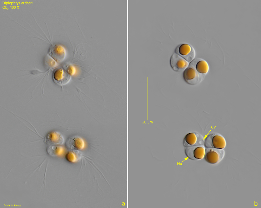
Sometimes specimens in cell division can be found (s. fig. 4 a-b). Depending on how often the mother cell divides, stages with 2, 4, or 8 cells can be found. If cell division continues to occur more frequently, spherical clusters of daughter cells form, which initially have a diameter of only 4–5 µm.
Fig. 1:Diplophrys archeri. D = 17.9 µm. A specimen with widely spreaded, branched pseudopodia. Nu = nucleus. Obj. 100 X.
Fig. 2 a-b:Diplophrys archeri. D = 11 µm. Two focal planes of a smaller specimen. CV = contractile vacuole, Nu = nucleus. Obj. 100 X.
Fig. 3:Diplophrys archeri. D = 15.6 µm. A second specimen with spreaded pseudopodia. Note the hyaline membrane (HM) covering the cell. Obj. 60 X.
Fig. 4 a-b:Diplophrys archeri. Two specimens in the process of cell division. CV = contractile vacuole, Nu = nucleus. Obj. 100 X.