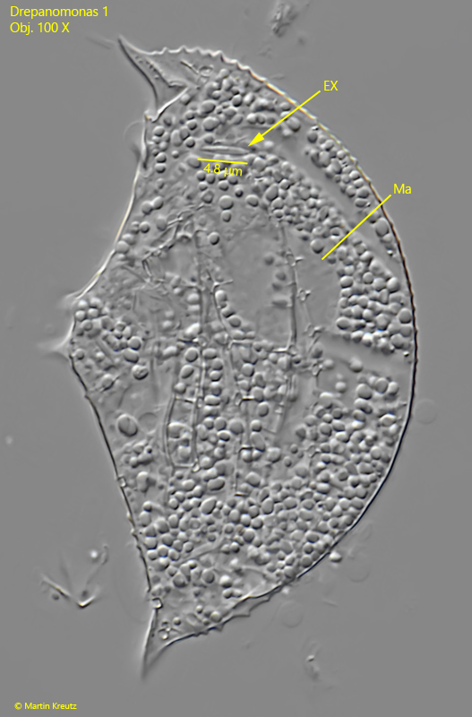
Drepanomonas 1
Most likely ID: Drepanomonas nov. spec.
Â
Synonym: n.a.
Â
Sampling location: Sima Moor (Austria)
Â
Phylogenetic tree: n.a.
Â
Diagnosis:Â
- body crescentic with tapered ends
- laterally flattened with lateral ribs
- in midbody a long, sharp main spine with a secondary spine
- one anterior spine on left side
- cortex with distinct pores with somatic cilia
- length about 70 µm
- cytostome in a depression in mid-body
- globular macronucleus in anterior half
- one spherical micronucleus, adjacent to macronucleus
- one contractile vacuoles near cytostome
- extrusomes spindle-shaped,about 5 µm long
- longitudinal ribs of cortex serrated
No drawings from previous authors available.
So far, I have only found one specimen of Drepanomonas 1 in June 2024 in the Sima Moor in Austria. Unfortunately, I was only able to examine the specimen from the left side.
Drepanomonas 1 has a prominent, long, and pointed spine about 8 µm in length on the ventral side in the middle of the body. Directly above it is a much smaller secondary spine. Additionally, near the front end on the left side, there is a further small spine. On the left side, three longitudinal ribs run along the body shape. They show distinct serration, as does the dorsal body margin. The mouth opening is located in the middle, and just below it is the contractile vacuole. The globular macronucleus in my specimen was located in the anterior half of the body. The small micronucleus was hard to see. Spindle-shaped extrusomes about 5 µm in length are embedded in the longitudinal ribs.
Fig. 1 a-d: Drepanomonas 1. L = 68 µm. A freely swimming specimen from left. Obj. 60 X.
Fig. 2 a-c: Drepanomonas 1. L = 68 µm. Different focal planes of a slightly squashed specimen from left. The mouth opening is a cavity near mid-body. EX = extrusome. Obj. 100 X.
Fig. 3 a-c: Drepanomonas 1. L = 68 µm. On the ventral side in mid-body a long and sharp main spine (MS) is visible with a small secondary spine (SS) above). Near the anterior end a small spine (AS) is visible in the left side. The contractile vacuole (CV) is located near the mouth opening. Mi = micronucleus. Obj. 100 X.
Fig. 4: Drepanomonas 1. In the strongly squashed specimen the globular macronucleus (Ma) is visible as well as the spindle-shaped extrusomes (EX) with a length of about 5 µm. Obj. 100 X.