oral bulge with bundle of 18–30 µm long, rod-shaped extrusomes
pellicle covered with mucilaginous sheath
macronucleus broadly ellipsoid
one micronucleus adjacent to macronucleus
contractile vacuole terminal
No drawings from previous authors available.
So far, I have only found one specimen of Enchelyodon 1 in January 2025 in the Mühlweiher Litzelstetten. The specimen was found among floating, dead aquatic plants. Since there were other objects under the coverslip, I was unfortunately unable to fix and squash the specimen for a more detailed examination.
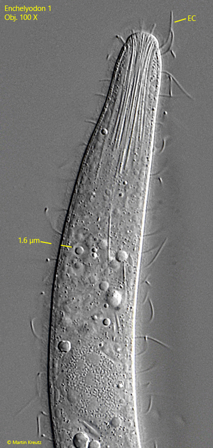
Due to the oral bulge with the shape of a flat, convex dome, I believe that this ciliate belongs to the genus Enchelyodon. The other characteristics also support this. The oral bulge surrounded by a ring of elongated cilia (s. fig. 3). Additionally, the pharynx is equipped with a dense bundle of extrusomes (s. fig. 2). These are thin, rod-shaped, and according to my measurements, between 18–30 µm long. The extrusomes are somewhat flexible but not curved or thickened at the ends. The body is covered by a delicate mucilaginous layer 1.5-2.0 µm thick, which is not unusual for the genus Enchelyodon (s. figs. 2 and 3). Probably, scales are also embedded in this mucilaginous layer, which cannot be resolved with a light microscope.
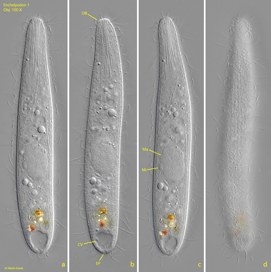
The body of Enchelyodon 1 is slender ellipsoid with nearly parallel sides. The macronucleus of my specimen was broadly ellipsoid with an attached micronucleus, the exact shape of which I could not discern (s. fig. 1 c). The contractile vacuole is terminal with a clearly visible excretory pore at the posterior pole (s. fig. 1 b). I could not detect the dorsal brush.
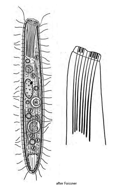
I could not find an Enchelyodon species with these characteristics in the literature available to me. The similar species Enchelys vestita is significantly larger at 200–220 µm in length and also has a distinctly kidney-shaped macronucleus. Another possibility to consider is Cataphractes austriacus (s. fig. 4). This species matches many features of my specimen, but not the shape and composition of the extrusomes. Cataphractes austriacus has two types of extrusomes (s. fig. 4). The larger type 1 are slightly curved with a length of 25–30 µm and a thickened end, while the type 2 extrusomes are only 2–3 µm long and rod-shaped. I could not recognize this form and composition of extrusomes in my specimen, which is why it might possibly be a previously undescribed, new species Enchelyodon nov. spec.
Fig. 1 a-d:Enchelyodon 1. L = 128 µm. A freely swimming specimen. CV = contractile vacuole, EP = excretion pore, Ma = macronucleus, Mi = micronucleus, OB = oral bulge. Obj. 100 X.
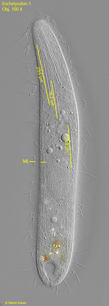
Fig. 2:Enchelyodon 1. L = 128 µm. Focal plane on the thin, rod-shaped extrusomes with a length of 18–30 µm. The body is covered by a decilate mucilaginous layer (ML). Obj. 100 X.
Fig. 3:Enchelyodon 1. A strongly contrasted image for visualization of the mucilaginous layer with a thickness of 1.6 µm. Obj. 100 X.
Fig. 4: The similar species Cataphractes austriacus after Foissner.